
Journal of Clinical Images and Medical Case Reports
ISSN 2766-7820
Case Report - Open Access, Volume 4
Pachychoroid and detachment of the
pigment epithelium: Case report
Akannour Younes*; El Akhdari Meryem; Louai Serghini; Abdallah Elhassan; Berraho Amina
Department of Ophthalmology B, Hospital of Specialties of Rabat (CHU Ibn Sina), Mohammed V University, Morocco.
*Corresponding Author : Akannour Younes
Service of Ophthalmology B, Hospital Specialties Rabat (IBN sina), University Mohammed V, Morocco.
Tel: +212676082240;
Email: younesah034@gmail.com
Received : Mar 20, 2023
Accepted : Apr 10, 2023
Published : Apr 17, 2023
Archived : www.jcimcr.org
Copyright : © Younes A (2023).
Abstract
The article discusses a case report of a 49-year-old patient who presented with an isolated pigment epithelium detachment. Multimodal imaging revealed the presence of a pachychoroid, leading to the diagnosis of a pachychoroid pigment epitheliopathy. Pachychoroidopathies are a recent description that includes choroidal thickening, choroidal hyperpermeability, and pigment epithelium alterations without a retinal serous detachment. The major risk associated with pachychoroid is the development of choroidal neovascularization, which requires close monitoring and treatment if necessary. In conclusion, when encountering isolated pigment epithelium detachment associated with pachychoroid in a young subject, it is necessary to consider the possibility of pachychoroid pigment epitheliopathy and closely monitor the patient for potential complications.
Keywords: Pachychoroid; Detachment of the pigment epithelium; Multimodal imaging.
Citation: Younes A, Meryem EA, Serghini L, Elhassan A, Amina B, et al. Pachychoroid and detachment of the pigment epithelium: Case report. J Clin Images Med Case Rep. 2023; 4(4): 2373.
Introduction
The detachment of the pigment epithelium corresponds to a separation between the pigment epithelium and the Bruch’s membrane. It can be associated with other clinical signs or isolated. The pachychoroid associates choroidal thickening and hyperpermeability with alterations of the pigment epithelium without the presence of a retinal serous detachment. We report the case of a 49-year-old patient presenting with an isolated pigment epithelium detachment, whose multimodal imaging led to the discovery of a pachychoroid, leading to the diagnosis of a pachychoroid pigment epitheliopathy.
Clinical case
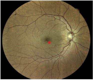
We report the case of a 49-year-old patient with no particular medical history who presented to an ophthalmology consultation for presbyopia optical correction. The ophthalmologic examination revealed visual acuity (with correction) of 10/10, P3 in the right eye and 10/10, P2 in the left eye. The anterior segment examination was unremarkable, while the fundus examination of the right eye revealed the presence of a bubble in the inferotemporal region of the fovea (Figure 1).
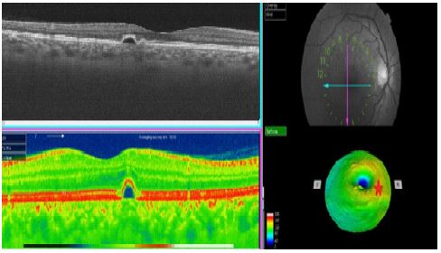
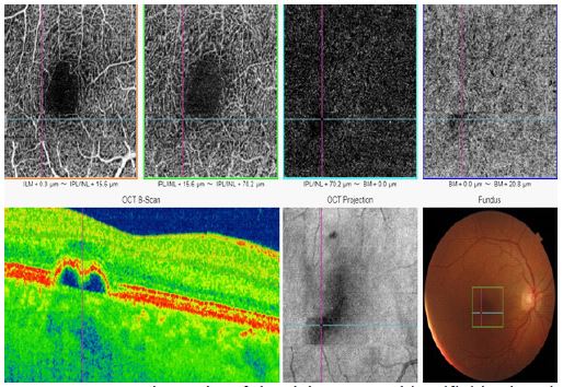
We decided to perform an OCT, which revealed the presence of a round hyporeflective homogenous content pigment epithelium detachment, without signs of exudation, located in the temporomacular region with a respected foveal depression. EDI mode allowed us to find a thickened choroid with dilated choroidal vessels. The presence of this pigment epithelium detachment prompted us to perform an OCT-A, which allowed us to rule out the presence of choroidal neovascularization. Given the isolated and harmless nature of the pigment epithelium detachment, without any signs of gravity identified by multimodal imaging, surveillance was recommended for the patient.
The diagnosis of a pachychoroid pigment epitheliopathy was made based on the association of an isolated pigment epithelium detachment with a pachychoroid in a young patient. A follow-up of 6 months did not reveal any functional or structural deterioration.
Discussion
The term pachychoroid comes from the ancient Greek “pachy,” which means thick, and refers to different pathologies that have in common a thick choroid. The detachment of the pigment epithelium occurs between the basal part of the pigment epithelium and the inner layer of the Bruch’s membrane [1]. It is better visualized by OCT and its content can be more or less reflective. In the vast majority of cases (> 60%), it is of degenerative origin associated with AMD (macular degeneration related to age) [2]. Isolated pigment epithelium detachments are rather rare.
Multimodal retinal imaging, including color retinography, autofluorescence and infrared images, spectral domain OCT (if possible in EDI or swept-source), fluorescein and indocyanine green angiography, and OCT-angiography are tools that provide arguments regarding the nature of the pigment epithelium detachment and form a bundle of arguments about their origin. The essential point is to distinguish neovascularized pigment epithelium detachments from other pigment epithelium detachments [3]. Pachychoroidopathies are a recent description dating back to 2013 by K. B. Freund [4]. They include choroidal thickening, choroidal hyperpermeability, and pigment epithelium alterations without a retinal serous detachment [4]. The pathophysiological mechanisms explaining pachychoroid are unknown. The best examination to assess choroidal thickness is OCT in Enhanced Deep Imaging (EDI) mode. There is a wide spectrum of pathologies associated with pachychoroid, which can be complicated by choroidal neovascularization [5]. Epitheliopathy pigmentosa pachychoroid is part of the clinical spectrum of anomalies with pachychoroid. It particularly affects patients around 50 years old with type A personality, and the involvement is often bilateral and asymmetric [6]. In addition to the pachychoroid detected by OCT-EDI, there are alterations of the pigment epithelium without the presence of subretinal fluid [4]. The major risk is the development of choroidal neovascularization [5], which requires close monitoring of these young patients to identify any functional or structural worsening that would then require treatment with IVT anti-VEGF.
Conclusion
When encountering isolated pigment epithelium detachment associated with pachychoroid in a young subject, it is necessary to consider the possibility of epitheliopathy pigmentosa pachychoroid. Close monitoring is required to detect neovascular complications requiring treatment with IVT anti-VEGF. In case of diagnostic doubt, close follow-up often allows detection of serious forms as well as benign forms.
Declarations
None of the authors has any conflicts of interests to disclose
Funding: The authors received no financial support for the research, authorship, and/or publication of this article
References
- Murphy RP, Yeo JH, Green WR, et al. Dehiscences of the pigment epithelium. Trans Am Ophthalmol Soc. 1985; 83: 63-81.
- Schmidt-Erfurth U, Waldstein Sm, Deak Gg, et al. Pigment epithelial detachment followed by retinal cystoid degeneration leads to vision loss in treatment of neovascular age-related macular degeneration. Ophthalmology. 2015; 122: 822-832.
- PAULEIKHOFF D, LÖFFERT D, SPITAL G et al. Pigment epithelial detachment in the elderly. Clinical differentiation, natural course and pathogenetic implications. Graefes Arch Clin ExpOphthalmol. 2002; 240:533-538.
- Warrow DJ, Hoang QV, Freund KB. Pachychoroid pigment epitheliopathy. Retina. 2013; 33: 1659-1672.
- Fung AT, Yannuzzi LA, Freund KB. Type 1 (sub-retinal pigment epithelial) neovascularization in central serous chorioretinopathy masquerading as neovascular age-related macular degeneration. Retina. 2012; 32: 1829-1837.
- Pang CE, Freund KB. Pachychoroid pigment epitheliopathymaymasquerade as acute retinal pigment epitheliitis. Invest Ophthalmol Vis Sci. 2014; 3: 111-115.