
Journal of Clinical Images and Medical Case Reports
ISSN 2766-7820
Clinical Image - Open Access, Volume 4
Double rim sign: Typical appearance of brain abscess
Abir Lemrabet*; El Haddad S; Iraqui Houssaini Z; Allai N; Chat L
Pediatric Teaching Hospital, Mohammed V University, Rabat, Morocco.
*Corresponding Author : Abir lemrabet
Pediatric Teaching Hospital, Mohammed V University, Rabat, Morocco.
Email: abirlemrabet8@gmail.com
Received : Mar 20, 2023
Accepted : Apr 11, 2023
Published : Apr 18, 2023
Archived : www.jcimcr.org
Copyright : © lemrabet A (2023).
Keywords: Brain abscess; Dual rim sign; MRI.
Citation: Lemrabet A, El Haddad S, Houssaini ZI, Allai N, Chat L. Double rim sign: Typical appearance of brain abscess. J Clin Images Med Case Rep. 2023; 4(4): 2376.
Description
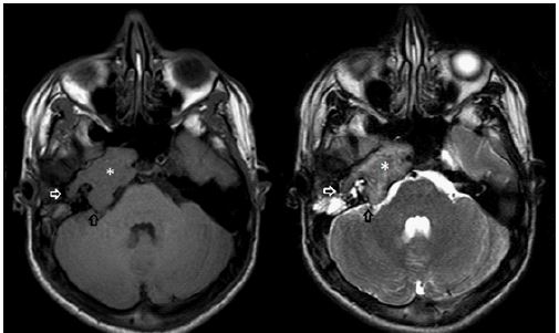
A 5-year-old child with no particular history consulted for tonic-clinical generalized seizures of recent onset, all evolving in a febrile context. The clinical examination revealed a slight disorientation, but there was no localized neurological deficit. An MRI was performed 72 h after the onset of the symptomatology. Cerebral masses were highlighted on this MRI in relation to cerebral abscesses (Figure 1).
Discussion
Cerebral abscesses are intracranial suppurations in a neoformed cavity, often insidious with a non-specific clinical presentation. They can develop by contiguity or by hematogenous route. The majority of cases of brain abscesses are polymicrobial, with a predominance of streptococci and strict anaerobes. In case of suspected brain abscess, brain imaging is necessary, with a preference for MRI. If this examination is not available within 24 hours, a CT scan is performed.
The characteristics of brain abscesses on MRI are well described; demonstrating a central portion appearing as T1 hyposignal/T2 hypersignal and a peripheral portion (capsule) appearing as T1 iso or hypersignal and T2/Flair hyposignal. Although peripheral enhancement of lesions is not specific to imaging, diffusion-weighted sequences showing central diffusion restriction are essential for the diagnosis of brain abscess (Figure 1) [1].
Another very discriminating sign of brain abscesses; the double rim sign; on susceptibility-weighted imaging (SWI) and T2, it consists of two concentric rims surrounding the central cavity, one of which is hypointense and the inner one relatively more hyperintense (Figure 1) [2].
The double rim sign is seen on MRI in 75% of brain abscesses and is very useful in differentiating an abscess from a necrotic tumor (glioblastoma, metastasis) [2].
References
- Leuthardt , EC , Wippold , FJ 2nd , Oswood , MC , Rich , KM. Imagerie IRM pondérée en diffusion dans l’évaluation préopératoire des abcès cérébraux. Chirurgie Neurol. 2002; 58: 395-402.
- Toh CH, Wei KC, Chang CN, et al. Différenciation des abcès cérébraux pyogéniques des glioblastomes nécrotiques à l’aide de l’imagerie pondérée en fonction de la sensibilité. AJNR Am J Neuroradiol. 2012; 33: 1534 - 1538.