Journal of Clinical Images and Medical Case Reports
ISSN 2766-7820
Clinical Image - Open Access, Volume 4
Acute epiglottitis in a young adult: Rare CT scan imaging
considering its life-threatening prognosis
Khadija El Bouhmadi*; Myriam Loudghiri; Youssef Oukessou; Sami Rouadi; Redallah Abada; Mohamed Roubal; Mohamed Mahtar
Department of Otorhinolaryngology, 20 August Hospital, Casablanca, Morocco.
*Corresponding Author : Khadija El Bouhmadi
Department of Otorhinolaryngology, 20 August Hospital, Casablanca, Morocco.
Email: elbouhmadik25@gmail.com
Received : Apr 03, 2023
Accepted : May 04, 2023
Published : May 11, 2023
Archived : www.jcimcr.org
Copyright : © El Bouhmadi K (2023).
Keywords: Epiglottitis; CT scan imaging; Emergency.
Citation: El Bouhmadi K, Loudghiri M, Oukessou Y, Rouadi S, Abada R, et al. Acute epiglottitis in a young adult: Rare CT scan imaging considering its life-threatening prognosis. J Clin Images Med Case Rep. 2023; 4(5): 2410.
Clinical image description
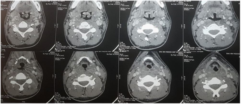
Acute epiglottitis is a life-threatening condition causing an inflammatory swelling of the epiglottis and nearby structures narrowing the upper airway that can lead to respiratory distress then arrest. The origin is often infectious, most frequently due to Haemophilus influenzae type B (HIB). More common in infants between 3 and 6 years, its paediatric incidence decreased thanks to vaccination while in the adult population, it remains stable. We received in our emergency department an eighteen years old man, with no pathological history, for acute moderate dyspnea with inspiratory stridor, a muffled voice and a severe dysphagia. We avoided examining the pharynx with a tongue blade since the manipulation of the oral cavity can raise the edema and aggravate the case. The diagnosis is primarily done on clinical suspicion but since the patient was still stable and the CT scan available, an imaging could have been done under close surveillance. The rarely obtained scan shows, as in our patient case, regular thickening and important edema of the epiglottis and the adjacent structures: arytenoids, aryepiglottic folds, and vallecula, tightening the airway, with a possible total obstruction. The prognosis depends on the rapidity of the medical care based on securing the airways. Neither intubation nor tracheostomy was needed for our patient, but he stayed under observation, receiving IV corticosteroids and antibiotics. The follow up was marked by clinical improvement and the ulterior examination with a flexible laryngoscope showed a back to normal anatomy.