
Journal of Clinical Images and Medical Case Reports
ISSN 2766-7820
Clinical Image - Open Access, Volume 4
Metastatic pancreatic carcinoma – the diagnosis
was on the skin
Teresa Medeiros1*; Raquel Machado-Neves2; Ana Oliveira3; Cristina Rosário1
1Serviço de Medicina Interna, Hospital Pedro Hispano, Unidade Local de Saúde de Matosinhos, Portugal.
2Serviço de Anatomia Patológica, Hospital Pedro Hispano, Unidade Local de Saúde de Matosinhos, Portugal.
3Serviço de Dermatologia, Hospital Pedro Hispano, Unidade Local de Saúde de Matosinhos, Portugal.
*Corresponding Author : Teresa Medeiros
Serviço de Medicina Interna, Hospital Pedro Hispano, Unidade Local de Saúde de Matosinhos, Portugal.
Tel: +35-1938361956;
Email: Teresa.pmedeiros@ulsm.min-saude.pt
Received : Apr 12, 2023
Accepted : May 11, 2023
Published : May 18, 2023
Archived : www.jcimcr.org
Copyright : © Medeiros T (2023).
Citation: Medeiros T, Neves RM, Oliveira A, Rosário C, et al. Metastatic pancreatic carcinoma – the diagnosis was on the skin. J Clin Images Med Case Rep. 2023; 4(5): 2419.
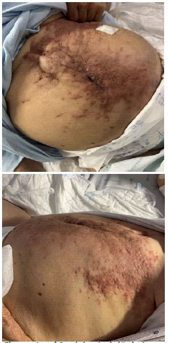
Description
A 82-year-old woman was admitted to the emergency room with dyspnea. On examination she had SaTO2 of 93% with FiO2 of 28%, crackles in the base of both lungs, edema of the lower limbs with godet and also nodular, erythematous, stone-like skin lesions, coalescing in the umbilical region that appeared 6 months before (Figures 1 and 2). She was applying topical medication without improvements. The patient had been diagnosed in the previous year with pancreatic ductal adenocarcinoma with vascular and duodenal invasion and underwent distal splenopancreatectomy and partial duodenectomy, but no systemic treatment.
Type 2 respiratory failure was documented, with right pleural effusion of moderate volume and increased Nt-proBNP (1289 ng/dL). The analysis revealed an amicrobial exudate with a predominance of mononuclear cells, with the cytological examination showing cells compatible with adenocarcinoma, with morphological and immunophenotypic aspects pointing to a primary pancreatic origin (Figures 3 and 4).
She was also evaluated by Dermatology which performed a skin biopsy that showed malignant epithelial cells suggestive of adenocarcinoma metastases and with immunohistochemical characteristics that also indicated a primary of pancreatic origin. It was decided not to treat the cancer. She died one month later.
Pancreatic cancer is the fourth leading cause of death from neoplastic etiology in the United States, accounting for 6% of oncological causes of death. Cutaneous metastases are uncommon, and, when present, they are located in the periumbilical area and favour disseminated disease, with a poor prognosis [1,2]. The dissemination mechanism is not well known, and several studies refer to direct invasion, local metastasis or hematogenous dissemination as the main paths [3]. In this clinical case, the mechanism seems to have been seeding after curative surgery, being a feared but common complication of surgery or diagnostic biopsy. Unfortunately, the patient spent months undergoing local topical treatment without improvement which delayed the diagnosis.

References
- Zhou HY, Wang XB, Gao F, Bu B, Zhang S, Wang Z, et al. Cutaneous metastasis from pancreatic cancer: A case report and systematic review of the literature. Oncol Lett. 2014; 8: 2654-2660.
- Abdelaziz M, Thorley D, Ahmed W. Cutaneous presentation of metastatic pancreatic cancer. J Surg Case Rep. 2022; 2022: rjac589.
- Otsuka I. Cutaneous Metastasis after Surgery, Injury, Lymphadenopathy, and Peritonitis: Possible Mechanisms. Int J Mol Sci. 2019; 20: 3286.