Journal of Clinical Images and Medical Case Reports
ISSN 2766-7820
Clinical Image - Open Access, Volume 4
Squamous cell carcinoma of the face with a focus of myiasis
Fauzia de Fátima Naime1,2*; Amanda Fortes Portela Ferreira1; Pedro Henrique Gutemberg Silveira1; Georges Antoine de Pinho Ishak1
1Instituto Paulista de Cancerologia, Department of Clinical Oncology, São Paulo, SP, Brazil.
2Conjunto Hospitalar do Mandaqui, Department of Clinical Medicine, São Paulo, SP, Brazil.
*Corresponding Author : Fauzia de F Naime
Instituto Paulista de Cancerologia, Department of Clinical Oncology, São Paulo, SP, Brazil.
Email: fauzianaime@yahoo.com.br
Received : May 08, 2023
Accepted : May 31, 2023
Published : Jun 07, 2023
Archived : www.jcimcr.org
Copyright : © Naime FF (2023).
Citation: Naime FF, Ferreira AFP, Silveira PHG, Pinho Ishak GA. Squamous cell carcinoma of the face with a focus of myiasis. J Clin Images Med Case Rep. 2023; 4(6): 2446.
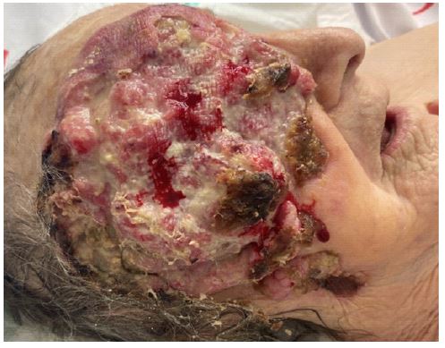
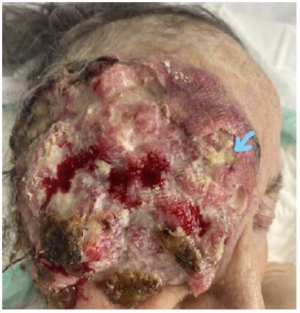
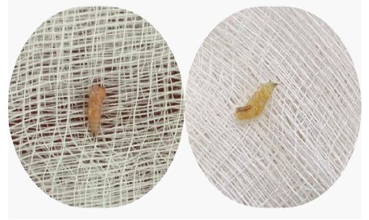
Clinical image description
An 87-year-old woman was admitted to a public hospital in São Paulo, Brazil, with an extensive, ulcerated, infected lesion with bleeding on her face, along with a focus of myiasis. The lesion had appeared 15 years ago on the right malar region, but the patient had never sought treatment. In the past year, the lesion had progressed more rapidly, involving the entire right hemiface and orbit region. Upon admission, local cleaning and removal of myiasis larvae were performed. Antibiotic therapy was initiated with ceftriaxone and clindamycin, but due to worsening of the local infection, meropenem and vancomycin was prescribed. The patient presented with significant bleeding from the lesion requiring blood transfusion. The CT scan of the face showed a lesion measuring 120 x 25 cm with involvement of deep tissues, signs of invasion of the masticatory muscles, erosion of the lateral wall of the orbit and ipsilateral zygomatic bone. Biopsy results revealed moderately differentiated invasive squamous cell carcinoma. The patient unfortunately passed away one week later.