
Journal of Clinical Images and Medical Case Reports
ISSN 2766-7820
Clinical Image - Open Access, Volume 4
The serpent or snake sign in liver hydatid cyst
Ramine Sboui; Mohamed Ali Chaouch*; Sadok Ben Jabra; Ghada Naghmouchi; Ibtissem Korbi; Faouzi Noomen
Department of Visceral and Digestive Surgery, Monastir University Hospital, Monastir University, Tunisia.
*Corresponding Author : Mohamed Ali Chaouch
Department of Visceral and Digestive Surgery, Monastir University Hospital, Monastir University, Tunisia.
Email: docmedalichaouch@gmail.com
Received : May 19, 2023
Accepted : Jun 06, 2023
Published : Jun 13, 2023
Archived : www.jcimcr.org
Copyright : © Chaouch MA (2023).
Citation: Sboui R, Chaouch MA, Jabra SB, Naghmouchi G, Korbi I, Noomen F. The Serpent or Snake Sign in Liver Hydatid Cyst. J Clin Images Med Case Rep. 2023; 4(6): 2454.
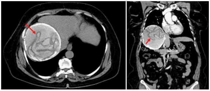
Clinical image description
The liver hydatid cyst is becoming a worldwide parasitic disease due to the immigrants from pandemic countries [1]. In some cases, the diagnosis is challenging and debatable and the imaging features are helpful. For this reason, the different radiological specific signs should be well known [2]. As an example, the serpent signs or snake signs is a radiological finding observed in liver hydatid cysts on imaging studies such as ultrasound, Computed Tomography (CT) or magnetic resonance imaging. It refers to the presence of daughter cysts, which are smaller cysts inside the larger cyst, in a serpentine or swirling pattern. This is due to the pressure difference between the larger cyst and the daughter cysts, causing the latter to move in a circulating manner. The serpent sign is considered a characteristic sign of hydatid disease and can aid in its diagnosis. We presented a CT-scan views of a 65-year-old women, with no particular past medical history, presenting a dysmorphic liver with a calcified cystic lesion at the expense of the right liver (segments V, VI, VII, VIII) with detachment of membrane realizing a serpent or snake sign, measuring 132 x 112 x 125 mm (Figure 1). Tt presents the following adhesions: supero-internal side with the IVC and the median side with the suprahepatic vein, infero-internal side with the common bile duct, portal vein and inferior vena cava, and in antero-inferior side with the gallbladder.
References
- Dziri C, Haouet K, Fingerhut A. Treatment of Hydatid Cyst of the Liver: Where Is the Evidence? World J Surg. août. 2004; 28: 731‑736.
- Chaouch MA, Dougaz MW, Cherni S, Nouira R. Daughter cyst sign in liver hydatid cyst. J Parasit Dis. déc. 2019; 43: 737‑738.