
Journal of Clinical Images and Medical Case Reports
ISSN 2766-7820
Clinical Image - Open Access, Volume 4
An uncommon encounter: A case report on developmental anomaly of the left lobe of the liver
*Corresponding Author : Shubhangi Yadav
Department of Anatomy, All India Institute of Medical Sciences- Raebareli, Uttar Pradesh, India.
Email: dr_shubhangi4@rediffmail.com
Received : May 20, 2023
Accepted : Jun 12, 2023
Published : Jun 19, 2023
Archived : www.jcimcr.org
Copyright : © Yadav S (2023).
Abstract
Developmental anomaly of liver are rare as compared to other abdominal viscera. With the advent of newer modalities of screening and the advancements in diagnosis and treatment, it is necessary that we should have a detailed knowledge of various morphological changes of left lobe of liver.
Citation: Yadav S. An uncommon encounter: A case report on developmental anomaly of the left lobe of the liver. J Clin Images Med Case Rep. 2023; 4(6): 2461.
Introduction
Normally the liver is divided into right and left lobes. In the initial development, the right and the left lobes are equal in size but due to the growth of neighboring organs on the left side, the left lobe regresses [1]. There can be congenital abnormalities of the liver such as agenesis of its lobe, deformed lobes, lobar atrophy, etc., [2]. Various studies have described extremely long left lobe - flat like a pancake, spatula-like, and lingular [3].
Case presentation
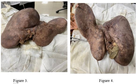
During the dissection of a 55-year-old female cadaver in the Department of Anatomy, AIIMS Raebareli, a large spatular prolongation of the left lobe of the liver was found. The left lobe was reaching the superior pole of the spleen and its width was more than that of the right lobe (Figures 1-4).

Discussion
The diagnosis of an elongated left lobe of the liver is established when the left lobe of the liver exceeds the stomach to the left and/or reaches the superior pole of the spleen on a CT image [4]. Various researchers have shown that this anomaly is associated with gastric volvulus and symptoms of pain, pressure, and pulling in the epigastric region.
Conclusion
The presence of the prolonged left lobe of the liver can be misinterpreted as splenic hematoma or tumor. These findings may be of great significance to surgeons and in USG and CT interpretations by radiologists.
References
- Veena vidya shankar, roopa kulkarni. Hypoplastic left lobe of the liver: a case report. Int j anat res. 2016; 4: 1958-1960.
- Aktan ZA, Savas R, Pinar Y, Arslan O. Lobe and Segment Anomalies of the Liver; J Anat. Soc. India. 2001; 50: 15-16.
- Dragica jurkovikj. New variant liver surface morphology according to portal vein segmentation: international journal of applied and pure science and agriculture Ijapsa. 2016; 2: 10-12.
- Size Wu, Rong Tu, Guangqing Liu, Ruixia Nan, Ying Guan, et al. Anatomical variation of the liver with elongated left lobe may be a trap for the ultrasound detection of focal liver lesion. Med Ultrason. 2015; 17: 12-15.