Journal of Clinical Images and Medical Case Reports
ISSN 2766-7820
Clinical Image - Open Access, Volume 4
Peritonitis after rosemary infusion: Is it possible?
Anwar Rahali*; Talha Laalou; Khawla Bahou; Said Benamer
Emergency Department, CHU Ibn Sina, Mohamed V University, Faculty of Medicine and Pharmacy, Rabat, Morocco.
*Corresponding Author : Anwar Rahali
Senior Resident, General Surgery, Department of Visceral Surgery II, Mohammed V Military Teaching Hospital, Mohamed V University, Faculty of Medicine and Pharmacy, Rabat, Morocco.
Email: rahali.anwar87@gmail.com
Received : May 12, 2023
Accepted : Jun 13, 2023
Published : Jun 20, 2023
Archived : www.jcimcr.org
Copyright : © Rahali A (2023).
Keywords: Peritonitis; Rosemary; CT scan; Diagnostic laparotomy.
Citation: Rahali A, Laalou T Bahou K, Benamer S. Peritonitis after rosemary infusion: Is it possible?. J Clin Images Med Case Rep. 2023; 4(6): 2463.
Background
We report a case of a 65-year-old-woman, with no pathological history. The patient developed acute onset of perpetual widespread abdominal pain and intermittent episodes of postprandial vomiting. She presented to the emergency department 24 hours after the beginning of symptoms. A physical examination revealed haemodynamically stable patient with generalized abdominal defense.
His baseline workup showed increased inflammatory markers levels such as plasma CRP and leukocytes.
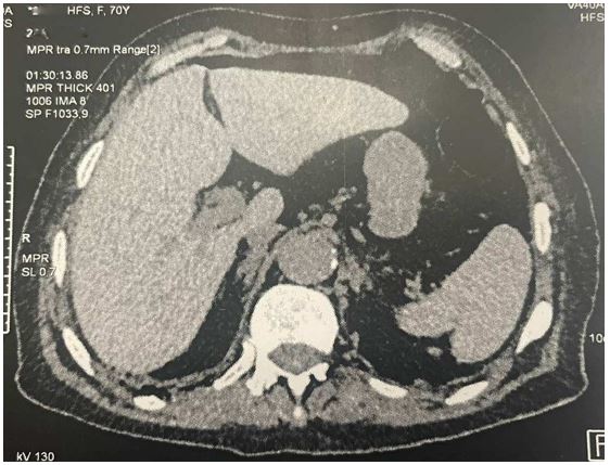
An abdominal contrast-enhanced CT showed perihepatic pneumoperitoneum and distended bowel loops; the findings were suggestive of peritonitis due to hollow visceral perforation (Figure 1).
Hence, a decision was made for a diagnostic laparotomy in which a punctiform bowel perforation caused by a dried rosemary stick was demonstrated (Figure 2). Then, removal of the stick, suture of the jejunal perforation, washing and drainage of the abdominal cavity were realized. The rest of the digestive tract appeared grossly unremarkable.
The patient was discharged on fourth postoperative day after complete oral feeding resumption. She was reviewed in consultation three months later, the clinical examination was unremarkable.