
Journal of Clinical Images and Medical Case Reports
ISSN 2766-7820
Clinical Image - Open Access, Volume 4
Apocrine cancer: Clinical image
*Corresponding Author : Laaliaoui Aymen
Department of Residence Janan Pasteur BV Abdelmoumen Casablanca, Morocco.
Email: aymen_laaliaoui@hotmail.com &
aymen_laaliaoui@gmail.fr
Received : May 29, 2023
Accepted : Jun 16, 2023
Published : Jun 23, 2023
Archived : www.jcimcr.org
Copyright : © Aymen L (2023).
Abstract
The incidence of apocrine carcinoma, a rare subtype of breast cancer, ranges from 0.3 to 4% [1]. Currently, its prognosis and treatment are the same as those of other ductal carcinomas without apocrine disruption [2,3]. However, the management of patients may be affected by its recognition.
Citation: Aymen L. Apocrine cancer: Clinical image. J Clin Images Med Case Rep. 2023; 4(6): 2469.
Description
We report a case of a 61-year-old woman sought our institution’s advice regarding an autopalpable nodule in her left breast. Breast retraction and inflammatory symptoms at the level of the left superexternal quadrant.
This patient lacked a family history of cancer
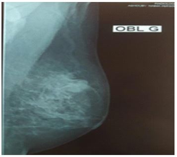
The patient underwent a mammogram, which revealed the presence of two overcrosses of contiguous circumscribed opacities in the left breast: One measuring 20 mm well limited with polylobed contours and the other measuring 15 mm poorly limited seat of a focus of fine, irregular microcalcifications with suspension grouping and a slight thickening of the QS skin.

Right breast without abnormality echographic complement
On the left breast: Presence of two contiguous formations at the level of the QSE: one slightly cystic hypoechogenic, well limited with polylobate contours, measuring 18 x 17 mm, the other very attenuating hypoechogenic with irregular limits measuring 21 x 17 mm, surrounded by a thick and irregular hyperechogenic halo.
Free axillary hollows.
Straight boobs without abnormality Examining BIRADS 5 to the left.
In addition, the patient was treated with a nodule-based microbiopsy that objectified an invasive ductal carcinoma with undifferentiated small cells and carcinomatous lymphangitis in the fats. With an IHC profile: Negative RH, Her2:0, 60% Ki67=Triple negative.
The patient received 6 cures of neo-adjuvant chemotherapy based on (3 AC60 + 3 pacli).
The clinical examination carried out in our institution was found in the left breast of an induration mass taking the upper quadrants, ulcerated, with exulceration facing the nipple.
Right breast: no palpable nodule.
Free axillary hollows.
The patient received a left mastectomy and a homolateral axillary scrub with anapath: INFILTRATING NOS-TYPE BREAST CARCINOMA , Grade SBR III , EV + -In situ component 15% -IHC=RH=negative, HER2=0 , ki67= 80%=Triple negative axillary clearance = 4N+/5N.
References
- Rouche, Julia Arfi, Mathieu, Marie-Christine, Canale, et al. Carcinome apocrine in situ du sein: corrélation anatomo-radiologique. Imagerie de la Femme. 2012; 22. 30-35.
- WADER, Jyotsna V., JAIN, Akash, BHOSALE, Suresh J., et al. Apocrine carcinoma of breast: a case report with review of the literature. Case reports in pathology. 2013; 2013.
- El Fouhi, Majdouline, Benider, Abdellatif, Gaetan, et al. Profil épidémiologique et anatomopathologique du cancer de sein au CHU Ibn Rochd, Casablanca. The Pan African Medical Journal. 2020; 37.