
Journal of Clinical Images and Medical Case Reports
ISSN 2766-7820
Case Report - Open Access, Volume 4
34-year-old female with acute onset quadriparesis and recurrent carpopedal spasm
Dnyanesh N Morkar; Akashdeep Singh*; Priyanka Patil
KLE Academy of Higher Education and Research, Jawaharlal Nehru Medical College, Belagavi, Karnataka, India.
*Corresponding Author : Akashdeep Singh
KLE Academy of Higher Education and Research, Jawaharlal Nehru Medical College, Belagavi, Karnataka, India.
Email: akashdp03@gmail.com
Received : May 22, 2023
Accepted : Jun 20, 2023
Published : Jun 27, 2023
Archived : www.jcimcr.org
Copyright : © Singh A (2023).
Keywords: Hypokalemic periodic paralysis; Gitelman syndrome; Carpopedal spasm.
Citation: Dnyanesh NM, Akashdeep S, Priyanka P. 34-year-old female with acute onset quadriparesis and recurrent carpopedal spasm. J Clin Images Med Case Rep. 2023; 4(6): 2473.
Background
Gitelman syndrome is an autosomal recessive condition also characterized by hypokalemic metabolic alkalosis, with hypocalciuria and hypomagnesemia. The similarity between the features of Gitelman syndrome and those caused by thiazide administration originally suggested that the defect might be in the distal convoluted tubule. The condition is linked to inactivating mutations in the gene for Na Cl- Cotransporter (NCCT). Loss of NCCT function results in Na+ and Cl- wasting from this segment, leading to hypovolemia with secondary activation of the renin-aldosterone system. However, the resulting increase in collecting tubule Na+ reabsorption is counterbalanced by K+ and H+ excretion, causing hypokalemic alkalosis.
Case presentation
A 34-year-old woman was brought to the emergency department by Emergency Medical Services (EMS) with complaint of Inability to get up from sitting position from past 1 day.
Patient was asymptomatic 1-day back when she went to wedding where she ate a lot of sweets and did strenuous exercise. After returning from wedding patient developed Fever and generalised body pain at 8:00 pm for which she went to local doctor and took medication. At 3:00 am patient developed weakness involving both upper and lower limb (Lower limb>Upper limb). Weakness was sudden in onset and did not progress. Patient could not get up from lying down position so she sat down and crawled down to reach the kitchen for drinking water where she was unable to hold the glass of water and glass fell down. Next morning she went to there family doctor with complaints of weakness in both upper and lower limb.
She was examined and was referred to KLE doctor Prabhakar kore hospital for further evaluation and management.
Weakness affected muscle of both upper and lower limb. Weakness was associated with Difficulty in wearing sleepers (can’t put fingers into sleepers), Difficulty while getting up from sitting position, Difficulty while sitting, Difficulty in combing hair, Difficulty in changing cloth, Difficulty in climbing stairs, Difficulty in performing hand grip.
No history of Tingling, Numbness, Back pain, swaying to either side, bowel bladder abnormality.
Diet H/o - patient ate lot of carbohydrates and sweet in the wedding she went.
Past history
Hypertensive * 4 years on T. Betacard AM (1-0-0).
H/o recurrent Cramps? Tetany * 2 years on T. Calcium(0-1-0).
H/o Similar episode of weakness * 7 month back resolved spontaneously and did not consulted any doctor for it.
Not a k/c/o Type 2 Diabetes Mellitus.
No H/o kidney disease.
No H/o drug intake like diuretics, statins.
Personal history
On Mixed diet.
Never consumed alcohol nor smoked cigarettes.
Family history
No history of cardiovascular, neurological or autoimmune diseases
On examination, pulse rate was 71 bpm, blood pressure 138/84 mm Hg, and respiratory rate was 14 /minute SpO2 99%. Patient was afebrile (36.4C) (Vitals within normal limits).
● CVS -S1 S2 heard
● Respiratory system - B/L air entry present
● Per abdomen - Soft and nontender
● CNS
Patient was conscious, alert and oriented in the emergency department.
Cranial nerve examination - Normal
Sensory system examination - Normal
Table 1: Motor system examination
| Left | Right | ||
|---|---|---|---|
| Power: | Upper limb | 5/5 | 5/5 |
| Lower limb | ⅘ | ⅘ | |
| Plantar: | Flexion | Flexion | |
| DTR: | Upper limb | +2 | +2 |
| Lower limb | +1 | +1 |
Pupil equal and reactive to light
Speech comprehensive
Cerebellum system examination- Normal
No nuchal rigidity.

Important laboratory investigations
| Day 1 4:50 pm | Day 1 10:45 pm | Day 2 9:00 am | Day 2 11:00 pm | Day 3 | Day 4 | Day 5 | Day 7 | |
|---|---|---|---|---|---|---|---|---|
| HB | 10.40 | 10.40 | 11.80 | |||||
| PLATELET | 262 | 262 | 220 | |||||
| TLC | 9.45 | 9.45 | 4.22 | |||||
| CALCIUM | 9.8 | 8.6 | 9.3 | 9.2 | ||||
| FREE CALCIUM | 0.99 | 1.00 | 1.05 | |||||
| UREA | 23 | 27 | 15 | |||||
| CREAT | 0.54 | 0.6 | 0.54 | |||||
| NA | 139 | 136 | 140 | 140 | 138 | |||
| K | 2.03 | 4.5 | 2.20 | 2.41 | 3.8 | |||
| MAGNESIUM | 1.8 | |||||||
| HCO3 | 12 | 30 | 29 | |||||
| CL | 99 | 99 | 100 | |||||
| T. BILI | 0.5 | 0.5 | ||||||
| D. BILI | 0.2 | 0.2 | ||||||
| SGOT | 26 | 26 | ||||||
| SGPT | 31 | 31 | ||||||
| ALP | 92 | 92 | ||||||
| T. PROTEIN | 7.1 | 7.1 | 4.3 | |||||
| ALBUMIN | 3.7 | 3.7 | 2.3 | 3.5 | ||||
| A:G | 1.1 | 1.1 | ||||||
| BUN | 10.74 | |||||||
| URIC ACID | 5.5 | 2.9 | 2.1 |
| Normal Value | Day 2 | Day 3 | Day 4 | |
|---|---|---|---|---|
| Urinary PH | 4.6-8.0 | 7.5 | ||
| Urinary Creatinine | 0.74-1.35 mg/dl | 48.39 mg/dl | ||
| Urinary Calcium | 100-300 mg/dl | 10.20 mg/dl | ||
| Urinary Calcium Creatinine Ratio | < 0.14 mg/dl | 0.19 mg/dl | ||
| Urinary Albumin | Negative | |||
| Urinary Sodium | 20 mEq/L | 141 mEq/L | 101 mEq/L | |
| Urinary Chloride | 110-250 mEq/d | 650 mEq/L |
S. MAGNESIUM LEVEL=1.5 mg/dl (normal 1.6-2.2) Day 2.
S.TS =1.86 mcIU/ml (normal=0.27-4.2) Day 2.
PTH=33.0 pg/ml (normal=15.0 -65.0) Day 2.
25-OH VITAMIN D=21.10 (normal=21-29) Day 2.
Spot urine potassium=29 mEq/L (normal =< 20 mEq/L) Day 3.
24-hour urine potassium=68.2 mEq/L (normal < 30 mEq/L) day 3.
Cpk -15 mcg/L (normal 10-20 mg/L) day 2.
Normal renal ultrasound with the absence of renal abnormalities or nephrocalcinosis day 2.
Peripheral smear - microcytic hypochromic with neutrophilic leukocytosis.
Urine culture sensitivity-negative
Day 1: Patient Serum electrolytes and ABG were sent along with all Routine investigations. Patient gave history of having meal rich in carbohydrate and involving in strenuous exercise leading to hypokalemia so most appropriate differential diagnosis would be? Hypokalemic periodic paralysis. Potassium came out to be 2.03 mEq/L and ABG showed metabolic alkalosis. Patient was started on IVF NS with 2 ampule of KCL @50 ml/hr and Syrup Kesol 10 ml in 50ml water (1-1-1).
Repeated Potassium in evening of day 1 came out to be 4.5 mEq/L.
Differential diagnosis
Hypokalemic periodic paralysis
Hyperkalemic periodic paralysis
Hypothyroidism
Hyperthyroidism
Hypoparathyroidism
Hyperparathyroidism
Glucocorticoid excess
Acromegaly
Diabetes mellitus
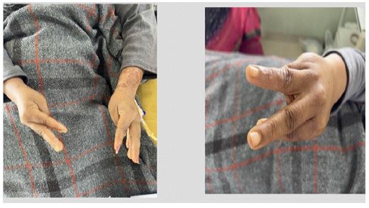
Day 2: Morning Potassium and Free calcium came out to be 2.20 mEq/L and 0.99 mmol/L respectively both on lower side. In evening patient developed CARPOPEDAL SPASM involving both hands and face. She was given 10% calcium gluconate 10 ml slowly over 10 min i.v. Calcium and free calcium of the patient were found to be 8.6 mg/dl and 1.00 mmol/L respectively. Free calcium being on lower side. Repeated MR also showed Potassium to be 2.41 mEq/L. So there was discussion on persistent drop of potassium.
Patient has No H/O low oral intake, Vomiting , Diarrhoea, Insulin history, Hypothermia, Steroid intake, Dialysis. All the causes that could lead to hypokalemia were ruled out. For low free calcium level, parathyroid level and Vitamin D level were found to be 33.0 pg/ml and 19.10 ng/ml respectively both being under Normal limits.
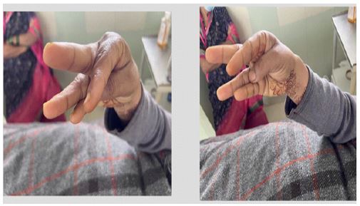
Day 3: Patient had another episode of carpopedal spasm involving face and both hands. Patient was given 10% calcium gluconate 10ml over 10 minutes i.v.
This episode lasted for 10 min.
Looking into consideration the loss of Potassium and Free calcium point towards Renal Loss. Renal tubular acidosis type 1 should have Non Anion gap metabolic acidosis but patient ABG showed metabolic alkalosis.
With renal loss of potassium and calcium through renal route patient was diagnosed as? Gitelman syndrome and was started on:
● T. Spironolactone 50 mg (1-0-1)
● T. Calcium, Phosphorus, Magnesium, Zinc And Vitamin D3 (1-0-1)
● Syrup Potassium Chloride 10 ml (1-1-1)
● Cap Calcitriol 0.25 mg (1-0-1)
On repeating free calcium and potassium it was found out to be 1.05 mmol/L and 4.98 mEq/L respectively.
Diagnosis-Hypokalemic Periodic Paralysis with Tetany Secondary to Gitelman Syndrome with Hypertension.
Discussion
Gitelman syndrome is an autosomal recessive condition also characterized by hypokalemic metabolic alkalosis, but with hypocalciuria and hypomagnesemia.
The similarity between the features of Gitelman syndrome and those caused by thiazide administration originally suggested that the defect might be in the distal convoluted tubule.The condition has now been linked to inactivating mutations in the gene for NCCT. Loss of NCCT function results in Na+ and Cl- wasting from this segment, leading to hypovolemia with secondary activation of the renin-aldosterone system. However, the resulting increase in collecting tubule Na+ reabsorption is counterbalanced by K+ and H+ excretion, causing hypokalemic alkalosis. The distal convoluted tubule normally reabsorbs only 7% to 8% of the filtered Na+ and Cl-load. The degree of volume contraction, the stimulation of the renin-angiotensin system, and the amount of K+ loss are therefore not substantial enough to stimulate PGE2 production. The hypocalciuria may be caused by enhanced proximal tubular calcium reabsorption, secondary to plasma volume contraction. The renal magnesium wasting is caused by downregulation of the epithelial magnesium channel TRPM6 in distal convoluted tubules.
In syndromes of chronic severe hypokalemia with metabolic alkalosis, the differential diagnosis may be facilitated greatly by taking into consideration the associated BP and the urinary chloride concentration.
Hypertension indicates disorders related to hyperaldosteronism. Extrarenal or renal Cl- and Na+ losses are the cause.
Extrarenal loss of sodium occurs in diarrhea, vomiting, or burns and is characterized by very low Cl- concentrations in the urine, often 1 mmol/l.
Renal loss of salt with high urinary Cl- and Na+ is typical of Bartter syndrome, Gitelman syndrome, and diuretic use.
The absence of hypomagnesemia and hypocalciuria will then argue against Gitelman syndrome. Genotyping is recommended to diagnose overlap of Bartter and Gitelman syndromes.
Clinical manifestations
More severely affected patients complain of Generalized and muscle weakness, Inability to work for extended periods, Salt craving and a preference for licorice, Cardiac disturbances, Muscle cramps, Tetany. Chondrocalcinosis of the knees does occur later in life, a result of hypomagnesemia.
Diagnosis
Laboratory evaluation will show Moderate hypomagnesemia, severe hypokalemia, Hypocalciuria, High urinary chloride concentration, Absence of thiazides from the urine, BP will be in the low-normal range, Genotyping should be performed in questionable or incomplete syndromes.
Treatment
There is no consensus on the best mode of treatment for Gitelman syndrome. Magnesium at 5 to 15 mmol/day (e.g., as magnesium oxide or aspartate). Potassium supplements are usually given to improve muscle weakness or cramps. However, dosing may be limited by diarrhea and abdominal discomfort. In exceptional cases, parenteral Mg2+ has been infused. Indomethacin, cyclooxygenase-2 (COX-2) inhibitors, and spironolactone are usually not helpful. The long-term prognosis for cardiac and renal function as well as for general health is good.
References
- Gopinath B, Chauhan N, Achappa B. Hypocalcaemia and hyponatraemia masquerading the diagnosis of Gitelman syndrome . BMJ Case Rep. 2019; 12: bcr-2018-227886.
- Gandhi K, Prasad D, Malhotra V, Agrawal D. Gitelman’s syndrome presenting with hypocalcemic tetany and hypokalemic periodic paralysis. Saudi J Kidney Dis Transpl. 2016; 27: 1026-1028.