
Journal of Clinical Images and Medical Case Reports
ISSN 2766-7820
Case Report - Open Access, Volume 4
Endometrial ossification: Unusual cause of chronic pelvic pain in low-resource settings
Samweli Ndulila1; Oscar Ottoman2; Richard Kiritta1; Edgar Ndaboine1; Simplice Harusha4; Rajab Kidenda3; Dismas Matovelo1*
1Catholic University of Health and Allied Sciences (CUHAS), Department of Obstetrics & Gynecology, Mwanza, Tanzania.
2Catholic University of Health and Allied Sciences (CUHAS), Department of Pathology, Mwanza, Tanzania.
3Catholic University of Health and Allied Sciences (CUHAS), Department of Radiology and Imaging Mwanza, Tanzania.
4Sengerema Designated District Hospital, Department of Obstetrics & Gynecology, Mwanza, Tanzania.
*Corresponding Author : Dismas Matovelo
Catholic University of Health and Allied Sciences (CUHAS), Department of Obstetrics & Gynecology, Mwanza, Tanzania.
Email: magonza@bugando.ac.tz
Received : Jun 09, 2023
Accepted : Jun 26, 2023
Published : Jul 03, 2023
Archived : www.jcimcr.org
Copyright : © Matovelo D (2023).
Abstract
Introduction: Endometrial ossification is a rare condition in which its actual etiology and pathogenesis is controversial and debatable however, the condition is usually related to secondary infertility after abortion and endometritis.
Case presentation: A 32-year-old para 2 living 1 lady who presented with long-standing intermittent sharp pelvic pain for seven years. She was a self-referral to BMC gynecology outpatient unit due to chronic pelvic pain. No prior history of intrauterine copper device insertion. Upon evaluation, she had normal ovulatory cycles. A pelvic x-ray could not show any spine deformities or any pelvic abnormalities. During transabdominal pelvic ultrasound imaging, a well-defined thick linear hyperechogenic structure with acoustic shadowing was seen in the endometrial cavity measuring 2 x 3 cm aligned to endometrial strips.
Ultrasound guided sharp dilatation & curettage (D&C) was performed by using ovum forceps in which we successfully removed a tubular structure measuring 2.3 by 3.4 cm with sharp ends deep in the endometrium. Histopathological analysis revealed trabecula and extracellular matrix osteocytes in keeping with endometrial ossification.
Conclusion: Endometrial ossification can be effectively treated with ultrasound guided dilation and curettage as an alternative to hysteroscopy.
Keywords: Endometrial ossification; Chronic pelvic pain; Ultrasound-guided dilatation; Curettage.
Citation: Ndulila S, Ottoman O, Kiritta R, Ndaboine E, Matovelo D, et al. Endometrial ossification: Unusual cause of chronic pelvic pain in low-resource settings. J Clin Images Med Case Rep. 2023; 4(7): 2482.
Introduction
Endometrial ossification is a rare condition affecting reproductive aged women characterized by presence of mature or immature bone tissue in the endometrium. The incidence of endometrial ossification is estimated to be 3/10,000 cases and often results from termination or spontaneous abortion that leads to either persistence of fetal bone or may follow true osseous metaplasia of endometrial tissue [1,2].
The most common presenting symptoms are secondary infertility in more than 80% of cases and rarely heavy menstrual bleeding and chronic pelvic pain [3]. There are controversies in the mechanism of endometrial ossification however, the widely accepted theory is the metaplasia of the stromal cells into osteoblastic cells that produce mature bone [4,5].
Hysteroscopy is the gold standard for diagnosis and treatment for endometrial ossification, with histopathological examination as an adjunct diagnostic aid to hysteroscopy [3,6]. However, in resource scarce setting, ultrasonography examination offers an alternative to hysteroscopy. Furthermore, the majority of cases resume their fertility sooner once the tissue had been removed. Moreover, saline infusion hysterography with Doppler studies may be performed to evaluate the stratum basalis and spinosum if there is a chance of fertility [7]. Here, we report a case of endometrial ossification in a woman presenting with chronic pelvic pain.
Case presentation
A 32-year-old woman, para 2 living 1 complained of lower abdominal pain for seven years. The pain was severe and came and went at regular intervals. Symptoms began two weeks after termination of her 3rd pregnancy at 29 weeks’ gestation in 2015. Without any regular pattern, the pain intensified with activity and decreased with rest. She was kept on multiple analgesics, but she remained in excruciating agony for quite some time.
She had her first vaginal birth successfully ten years back in 2010, she then conceived again in 2014 in which it ended up with spontaneous abortion at 2 months without any adverse event reported thereafter. Prior to her third pregnancy, she continued to experience regular menstrual cycles, normal volume and length, her cervical cancer screening was negative, and she had never experienced symptoms suggestive of sexual transmitted infections.
Her ongoing suffering led to bouts of chronic tension and worry, as well as social withdrawal and marital problems, all of which contributed to divorce in 2019. In 2018 and 2019, she visited several different medical facilities and had two uterine evacuations, but the pain continued despite these procedures. In 2021, she went to three further Primary Healthcare (PHC) facilities and underwent a number of ultrasounds, all of which produced results that were inconclusive. After some time, she made the decision to check in at our medical center with above history.
Clinical examination revealed a young woman who appeared stressed but well-kept and well-nourished. She was not pale, her blood pressure was 111/65 mmHg, pulse rate 72 beats per minute, respiratory rate 18 cycles per minute and temperature of 36 Celsius, and normal oxygen saturation.
Her cardiovascular and respiratory findings were normal. Her abdomen was normal in contour and shape, with a tender hypogastrium, with no palpable mass. Her uterus appeared slightly bulky on bimanual examination with a positive cervical excitation test however on visual inspection vulva, vaginal and cervix were normal.
Investigations
She had serum βhCG of 0.3 miu/ml, her full blood count was within normal reference, Hemoglobin level was 12.4 g/dl, WBC 4 x 10⁹ Platelets count 234 x 103 and normal white blood cell differentials. She underwent trans-abdominal pelvic ultrasound, which revealed a well-defined thick linear hyperechogenic structure with acoustic shadowing in the endometrial cavity measuring 2 x 3 cm (Figure 1). Her pelvic x-ray was normal.
Transabdominal ultrasound findings
Management and follow-up: She had been scheduled for a dilatation and curettage (D&C), and with the help of grayscale 2D ultrasound guidance, a tubular structure that had been deeply adhered to the endometrium was evacuated. The tissue biopsy was sent to histopathology for analysis, and the patient was released the next day in excellent condition.

Histopathological findings
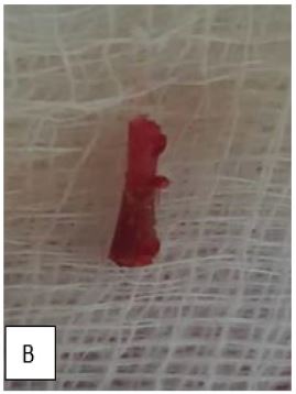
Gross findings: Showed multiple hemorrhagic tissue biopsy from endometrial curettage measured (2 x 1) cm mixed with hard calcified bone fragments measured 2 cm long as shown in figure 2 below.
Microscopic findings: Section showing fragments composed of endometrial gland and its stroma accumulated with area of bone trabecular some with calcification as shown in figures 3and 4 below.


Discussion
Endometria ossification is an occasional phenomenon mostly occurring following termination or spontaneous abortion in more than 80% of the cases reported [2]. About 100 cases worldwide have been documented. Its presentation varies but the frequent complaint is secondary infertility and heavy menstrual bleeding, and chronic pelvic pain [3,5].
Controversies exists in the mechanism of endometrial ossification; the widely accepted mechanism is metaplasia of the stromal cells into osteoblastic cells that produce mature bone. The endometrial ossification can be confused with an intra-uterine copper devices making difficult to diagnose [5,8].
Our patient presented with history of chronic pelvic pain for seven years with multiple uterine evacuation without conclusive results. However, prior to this, she had termination of an advanced pregnancy. This patient had termination of pregnancy at 29 weeks followed by 2 episodes of uterine evacuations. Upon review of literature, the major risk factor for endometrial ossification is the termination of pregnancy at least ≥ 3 months either spontaneous or surgical termination [3,9].
There was a delay in making of the diagnosis to this patient from lower health facilities due to lack of pathologists in her residential area and lack of clinician awareness of this condition [5]. On other hand our patient presented with unique and unusual activity-related pain which is an occasional symptom in endometrial ossification [10]. Infertility was not a concern for her because she had early been divorced. In several cases, diagnosis of endometrial ossification can be delayed due to the confusion with other diagnoses such as the Intrauterine Copper Devices (IUCDs), foreign bodies, calcified submucous fibroids, endometrial tuberculosis, Asherman’s syndrome, and rarities such as heterotopic bone and uterine malignant mixed Mullerian tumor and furthermore due to the fact that in most cases is silent unless it present with either infertility or abnormal uterine bleeding. In majority of cases, diagnosis of the endometrial ossification requires a high degree of suspicion, expertise and technology [5,11].
Endometrial ossification is best simultaneously diagnosed and treated with office hysteroscopy. Endometrium hypertrophy like white coral is usually visualized during hysteroscopy [3]. Hysteroscope offers the best modality in both diagnosing and removing the bone fragment [3,5,12], however, in resource limited setting like ours due to lack of hysteroscope we opted initially to perform D&C using an ovum forceps, but the bone could not be felt. Transvaginal ultrasound which is available in limited resource settings is an excellent alternative, with a high sensitivity and specificity for endometrial pathology, as applied to our patient [1].
After the removal of the bone from the endometrial cavity, the patient reported disappearance of the pelvic pains she had. The pain relief, resumption of fertility and less likelihood of recurrence after treatment has made it to be among diseases with good prognosis [3,5]. Furthermore, there is no role of hysterectomy in the management of endometrial ossification since most cases present in the reproductive age group and with infertility. Hysterectomy may be an option in women within perimenopausal group and have completed family [5].
Our case was different as the blind ovum forceps use was not successful, we improvised with ultra-sound guided with slight curette and successful removed the bone piece. On our side, we declare that this is the first time we encountered the case, and we present ultrasound guided D&C as an alternative to office hysteroscopy [3,13].
Conclusion
Ultrasound guided dilatation and curettage can be as effective as office hysteroscopy in treating endometrial ossification in low-resource settings.
Declarations
Patient’s perspective: The care provided was timely with full explanation of the diagnosis and prognosis and with a follow-up plan explained.
Acknowledgments: We are humbly grateful for the support and encouragement given by both Obstetrics and Gynecology department at Bugando Medical Centre and Catholic University of Health & Allied Sciences (CUHAS).
Timeline: The patient was admitted after the clinical workout, emergency surgery and other management were immediately performed to the patient. Preparation and completion of the case took 1 month, including follow up and after obtaining consent.
Author’s contribution: SN and DM played equal roles in evaluating the patient before surgery, performed surgery and prepared the initial drafts of this case report. OO did the histopathological studies of the sample and reviewed several draft of this manuscript, RK, SH and EN followed the patient post-surgery and later reviewed the final draft of the manuscript. RK did a pelvic ultrasound and later reviewed several drafts of the manuscripts. All authors read and approved the final manuscript.
Funding: The cost of care offered to this patient was partly covered by the patient and some was waived by the hospital administration. The cost of preparing this manuscript and publication was covered by the authors and the Catholic University of Health and Allied Sciences (CUHAS).
Consent for publication: Written informed consent was obtained from the patient for publication of this case and any accompanying images. A copy of the written consent is available for review by the Editor-in-Chief of this journal. Additionally, consent was sought and granted by the Catholic University of Health and Allied Sciences Directorate of Research and Publication to publish this work. A copy of the clearance document is also available for review by the Editor-in-Chief of this journal.
Competing interests: The authors declare that they have no competing interests.
References
- Poddar P, Chavan K, Saraogi RM, Yadav P. Endometrial Ossification: An Unusual Cause of Heavy Menstrual Bleeding (HMB). Journal of obstetrics and gynaecology of India. 2016; 66(Suppl 2): 666--668.
- Alorini M, Aziz M, Gromez A, Piton N, Sabourin JC. [Endometrial osseous metaplasia: A case report]. Annales de pathologie. 2017; 37(6): 488-490.
- Horo GA, Aka KE, Toure A, Koffi A, Seni K, Kone Met al. Endoscopy Management of Endometrial Ossification Associated With Secondary Infertility: A Case Report and Review of Literature. Journal of Clinical Gynecology and Obstetrics. 2016; 5(1): 45-49.
- Bahçeci M, Demirel LC. Osseous metaplasia of the endometrium: Aa rare cause of infertility and its hysteroscopic management. Human reproduction (Oxford, England). 1996; 11(11): 2537-2539.
- Nigar A, Yadav YK, Hakim S. Endometrial osseous metaplasia—-A rare presentation of polymenorrhagia: A case report. Journal Of Clinical And Diagnostic Research: JCDR. 2015; 9(4): QD07.
- Rosa ESJC, Barcelos ID, Navarro PA, Rosa ESAC, Nogueira AA, Ferriani RAet al. Osseous metaplasia of the endometrium associated with infertility: a case report and review of the literature. Journal of medical case reports. 2009;3:7427.
- Lainas T, Zorzovilis I, Petsas G, Alexopoulou E, Lainas G, Ioakimidis Tet al. Osseous metaplasia: Ccase report and review. Fertility and sterility. 2004; 82(5): 1433-1435.
- Gulec UK, Parlakgumus HA, Kiliçdag EB, Bolat F, Bagis T. Osseous metaplasia of the endometrium. BMJ case reports. 2010; 2010.
- Wani AH, Parry AH, Feroz I, Jehangir M, Rashid M. Imaging of endometrial osseous metaplasia—-an uncommon but treatable cause of infertility. Middle East Fertility Society Journal. 2020; 25(1): 35.
- Torné A, Jou P, Pagano R, Sanchez I, Ordi J, Vanrell JAet al. Endometrial ossification successfully treated by hysteroscopic resection. European journal of obstetrics, gynecology, and reproductive biology. 1996; 66(1): 75-77.
- Tsai MC, Arunamata A, Tristan S, Randall HW. Endometrial osseous metaplasia mimicking retained intrauterine device: a case report. The Journal of reproductive medicine. 2008; 53(11): 877-880.
- Amodeo S, Iannone V, Borriello M, Giambanco L. Hysteroscopic Management of Endometrial Osseous Metaplasia Mimicking a Foreign Body. Journal of minimally invasive gynecology. 2021; 28(10): 1673-1674.
- Coccia ME, Becattini C, Bracco GL, Scarselli G. Ultrasound-guided hysteroscopic management of endometrial osseous metaplasia. Ultrasound in obstetrics & gynecology : Tthe official journal of the International Society of Ultrasound in Obstetrics and Gynecology. 1996; 8(2): 134-136.