
Journal of Clinical Images and Medical Case Reports
ISSN 2766-7820
Clinical Image - Open Access, Volume 4
Hyalinosis cutis ET mucosae
Pratika Goya1; Dilip Meena2*
1Senior Resident, Department of Dermatology, Venereology and Leprosy, All India Institute of Medical Sciences, Bathinda-151001, India.
2Associate Professor, Department of Dermatology, Venereology and Leprosy, Teerthanker Mahaveer Medical College and Research Centre, Moradabad, Uttar Pradesh-244001, India.
*Corresponding Author : Dilip Meena, MD
Associate Professor, Department of Dermatology, Venereology and Leprosy, Teerthanker Mahaveer Medical College and Research Centre, Moradabad, Uttar Pradesh-244001, India.
Ph: +91-8191811380;
Email: dilip.aiims@gmail.com
Received : Jun 16, 2023
Accepted : Jun 29, 2023
Published : Jul 06, 2023
Archived : www.jcimcr.org
Copyright : © Meena D (2023).
Citation: Goyal P, Meena D. Hyalinosis cutis ET mucosae. J Clin Images Med Case Rep. 2023; 4(7): 2487.
Clinical image description
A 16-year-old male was brought to the Dermatology OPD with complaints of generalized progressive thickening of the skin since one year of life and development of blisters after trauma. There was a history of difficulty in swallowing, protrusion of tongue, hoarseness of voice, and diffuse loss of hair. Similar findings were also present in the younger sister. Cutaneous examination revealed the presence of multiple, yellowish, non-pruritic, infiltrated waxy papules involving the entire body with interspersed areas of non-tender hemorrhagic crusting and pox-like scarring, especially on the trunk (Figure 1). Hyperpigmented, verrucous plaques were present over bilateral elbows. There was a characteristic presence of beaded papules over the bilateral upper eyelid margins (Figure 2). Oral examination revealed the presence of macroglossia with lateral crenation, fissured oral commissures (Figure 3), and whitish waxy plaque over the left buccal mucosa (Figure 4). Histopathological examination revealed acanthotic epidermis with the deposition of eosinophilic, amorphous hyaline material in the papillary dermis and perivascular areas (Figure 5). The parents were counselled regarding the course and progression of the disease.
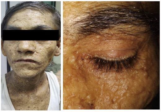
Figure 2: Characteristic beading of eyelid margins.


