
Journal of Clinical Images and Medical Case Reports
ISSN 2766-7820
Short Report - Open Access, Volume 4
Microphthalmos with elongated ciliary processes
Sarath S*; Rahul P Vijayakumar
Dr Rajendra Prasad Centre for Ophthalmic Sciences, All India Institute of Medical Sciences, New Delhi, India.
*Corresponding Author : Sarath S
Dr Rajendra Prasad Centre for Ophthalmic Sciences, AIIMS, New Delhi, India.
Tel: +919846113351
Email: sarathachu66@gmail.com
Received : Jun 23, 2023
Accepted : Jul 10, 2023
Published : Jul 17, 2023
Archived : www.jcimcr.org
Copyright : © Sarath S (2023).
Abstract
A 12-year-old female patient came to our outpatient department with complaints of low vision in both eyes since childhood. There was no significant history of ocular trauma or family history of similar disease. Her parents also give no significant medical or surgical history in the past. On examination her unaided vision was 3/60 in both eyes with manifest nystagmus. Slit lamp examination revealed bilateral small sized globe with smaller corneal diameters with prolonged ciliary processes attached to a partially subluxated lens. The superior ciliary processes were more elongated compared to the inferior ciliary processes resulting in inferior subluxation of the lens. She was referred to our low vision rehabilitation clinic for further management as there was no improvement of vision with glasses.
Keywords: Ciliary process anomaly; Anterior segment dysgenesis; Subluxated cataract; Congenital anomaly.
Citation: Sarath S, Vijayakumar RP. Microphthalmos with elongated ciliary processes. J Clin Images Med Case Rep. 2023; 4(7): 2502.
Introduction
Anterior segment dysgenesis refers to anatomical anomalies of the anterior segment structures such as cornea, iris, ciliary body, ciliary processes, and lens. These anomalies show vast phenotypic and genetic heterogeneity [1]. These includes Posterior embryotoxon, Axenfeld-Rieger syndrome, Peters anomaly, primary congenital glaucoma, Aniridia, Posterior Polymorphous Corneal Dystrophy, Iridocorneal Endothelial Syndrome, Iridogoniodysgenesis syndrome etc. Ciliary processes form a frill behind the iris around the lens margin in a circular manner [2]. There are around 60-80 ciliary process of which larger one measures around 2.5 mm only. They are continuous with the periphery of the iris in the front and the posterior surfaces relate to the suspensory ligament of the lens [3].
Case presentation
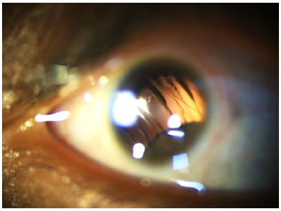
A 12-year-old female patient came to our outpatient department with complaints of low vision in both eyes since childhood with jerky eye movements noted by parents. The was no significant history of ocular trauma or family history of similar disease. Her parents also give no significant medical or surgical history in the past. On examination her unaided vision was 3/60 in both eyes with no further improvement with pinhole. Intraocular pressure was not recorded due to severe nystagmus on non-contact tonometry. She had manifest nystagmus with no head tilt. Extraocular muscle movements were normal with mild alternating convergent squint. Slit lamp examination revealed bilateral small sized globe with normal corneal shape and smaller corneal diameters of 6 mm in both eyes. Anterior chamber was deep with prolonged ciliary processes of approximately 5 mm more on the superior side attached to a partially subluxated lens. The superior ciliary processes were more elongated compared to the inferior ciliary processes resulting in inferior subluxation of the lens. The lens was clear but not covering the visual axis. The posterior segment ultrasound scan revealed a small lens in the anterior vitreous with an attached retina. A diagnosis of both eyes anterior segment dysgenesis with microphthalmos and microcornea with elongated ciliary processes and partially subluxated crystalline lens with manifest nystagmus was made. Considering the prolonged history of poor vision with lens subluxation and small sized globe we proceeded to manage her conservatively with visual rehabilitation and follow up to look for increase in subluxation or development of cataract. She was referred to our low vision rehabilitation clinic for further management as there was no improvement of vision with glasses.
Discussion
Anterior segment dysgenesis are rare developmental anomalies with a spectrum of conditions ranging from minor anomalies to major anomalies resulting in blindness since birth. Most of these are inherited with positive family history. In our case we could not find any positive family history. Elongated ciliary processes has not been described in the literature as a specific entity but few case reports shows that it can be associated with Persistent Hyperplastic Primary Vitreous (PHPV) or Persistent Foetal Vasculature (PFV) [4]. In our case the posterior segment ultrasound is anechoic with no foetal vasculature detected in the vitreous cavity. Also, the child had microphthalmia with microcornea and nystagmus which is bilateral. PFV presents unilaterally in most cases with or without microphthalmia or microcornea. Hence, we describe a distinct entity of anterior segment dysgenesis or anomaly with bilateral elongated ciliary processes along with microphthalmos and microcornea with nystagmus. Systemic examination also did not reveal any abnormality and hence we cannot call it a syndrome. Genetic studies and further evaluation can find out the inheritance pattern and help us to widen our knowledge on this new entity which we have described.
Declarations
Authors contributions: Sarath S was involved in patient evaluation and management, concept of case report, preparation of manuscript and final drafting.
Rahul P Vijayakumar was involved in image acquisition and editing of article.
Patient consent: The authors certify that they have obtained all appropriate patient and parents’ consent forms. In the form the patient and parents has/have given his/her/their consent for his/her/their images and other clinical information to be reported in the journal. The patient and parents understand that the patient’s name and initials will not be published and due efforts will be made to conceal their identity and maintain anonymity.
Conflict of Interest: Nil
Source of Funding: Nil
Acknowledgements: Nil
References
- Ito YA, Walter MA. Genomics and anterior segment dysgenesis: A review. Clin Experiment Ophthalmol. 2014; 42: 13-24.
- Smelser GK. Electron microscopy of a typical epithelial cell and of the normal human ciliary processTrans Am Acad Ophthalmol Otolaryngol. 1966; 70: 738.
- Delamere NA. Ciliary Body and Ciliary Epithelium. Adv Organ Biol. 2005; 10: 127-148.
- Warren N, Trivedi RH, Wilson ME. Persistent Fetal Vasculature with Elongated Ciliary Processes in Children. Am J Ophthalmol. 2019; 198: 25-29.