
Journal of Clinical Images and Medical Case Reports
ISSN 2766-7820
Clinical Image - Open Access, Volume 4
Don’t forget about me: Complications from a 40 year dropped gallstone
*Corresponding Author : Victor KO Chang
School of Medicine and Dentistry, Griffith
University, Gold Coast, Queensland, Australia.
Email: victorchang91@hotmail.com
Received : Jun 09, 2023
Accepted : Jul 12, 2023
Published : Jul 19, 2023
Archived : www.jcimcr.org
Copyright : © Chang VKO (2023).
Citation: Chang VKO. Don’t forget about me: Complications from a 40 year dropped gallstone. J Clin Images Med Case Rep. 2023; 4(7): 2506.
Description
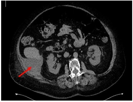
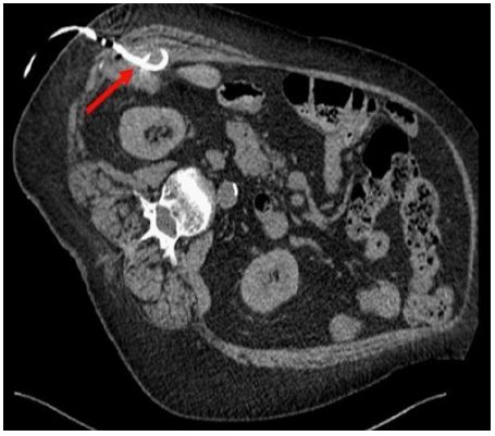
A 73-year-old Caucasian woman presented with a week’s history of vague post-prandial right upper quadrant and flank pain. She reported that the pain felt similar to her cholecystitis over 40 years ago for which she received a laparoscopic cholecystectomy (LC). She denied any other symptoms. She was haemodynamically stable and afebrile. Physical examination revealed only minor tenderness on palpation of her right flank with no evidence of peritonism. A CT abdomen was conducted revealing a 62 x 86 x 98 mm mixed density lesion in the retroperitoneum involving the transverse abdominus and internal oblique muscles, initially thought to represent a soft tissue tumour (Figure 1). Closer inspection revealed a well-defined 18mm rounded calcific focus at the inferior aspect of the lesion was consistent with retained gallstone (Figure 2) and associated abscess and granulomatous formation. Through the use of interventional radiology (IR), a percutaneous 8F drain was inserted (Figure 3) to decompress the abscess with subsequent stone removal.
LC, which is considered the gold standard treatment for cholecystitis, is commonly associated with spillage or dropped gallstones (DG), estimated in 2.3-40% of procedures [1]. DG harbours significant bacteria that acts as a nidus for infection. While most retained stones have no complications and patients remain asymptomatic, around 0.04% to 19% of cases leads to complications such as fistulas, granulomas, complications to the thoracic and reproductive systems, but intra-abdominal abscesses are by far the most common [1]. The average time to presentation is in the order of 4-5 months with the longest latency period of 20 years in documented literature [2,3]. Dropped gallstones are a pitfall for radiologists reviewing the images as they can imitate other pathology such as intra-abdominal neoplasia, peritoneal metastases, lymph nodes, focal liver masses and endometriosis [4]. In patients who have had laparoscopic cholecystectomies, dropped gallstones should be a differential diagnosis to consider irrespective of the temporal remoteness of the procedure.

References
- Jabbari Nooghabi A, Hassanpour M, Jangjoo A. Consequences of Lost Gallstones During Laparoscopic Cholecystectomy: A Review Article. Surgical laparoscopy, endoscopy & percutaneous techniques. 2016; 26(3): 183-92.
- Brueggemeyer MT, Saba AK, Thibodeaux LC. Abscess formation following spilled gallstones during laparoscopic cholecystectomy. JSLS : Journal of the Society of Laparoendoscopic Surgeons. 1997; 1(2): 145-52.
- Röthlin MA, Schöb O, Schlumpf R, Largiadèr F. Stones spilled during cholecystectomy: a long-term liability for the patient. Surgical laparoscopy & endoscopy. 1997; 7(5): 432-4.
- Ramamurthy NK, Rudralingam V, Martin DF, Galloway SW, Sukumar SA. Out of Sight but Kept in Mind: Complications and Imitations of Dropped Gallstones. American Journal of Roentgenology. 2013; 200(6): 1244-53.