
Journal of Clinical Images and Medical Case Reports
ISSN 2766-7820
Short Report - Open Access, Volume 4
A rare case of acute abdomen: Omental infarct
*Corresponding Author : Mustafa Salis
Department of General Surgery, Eskişehir City Hospital, Turkey.
Tel: +90-5383043317;
Email: salismustafa@gmail.com
Received : Jul 03, 2023
Accepted : Jul 20, 2023
Published : Jul 27, 2023
Archived : www.jcimcr.org
Copyright : © Mustafa S (2023).
Abstract
Omentum infarction occurs when the omentum is torsioned around its long axial axis and compresses the vascular structure. Although omentum infarction is rare, it is an important pathology that should be considered in acute abdomen. The insidence is 2 times higher in men than in women. Also more common in obese individuals. The pain usually localized in the right lower quadrant. Moderate leukocytosis, fever and vomiting may accompany the disease. Although primary excision of the infarcted area is accepted as the ideal treatment in the literature, in this case we decided to perform conservative treatment considering the omentum, which is called abdominal police, would limit the infarcted area. However, because the patients clinic did not regress, the patient was operated and the infarcted area was excised.
Keywords: Omentum infarct; Acute abdomen.
Citation: Mustafa S. A rare case of acute abdomen: Omental infarct. J Clin Images Med Case Rep. 2023; 4(7): 2517.
Introduction
Although omental infarction is rare, it is a pathology that should be kept in mind that can lead to acute abdomen. In the clinical differential diagnosis, conditions requiring emergency intervention such as acute cholecystitis, acute appendicitis, acute diverticulitis, renal colic, colon carcinoma perforation are included [1]. Although it is a condition that can be easily diagnosed with computed tomography, in many cases it can be diagnosed during surgery. Although the ideal treatment method in the literature is accepted as primary excision of the infarcted area, in our case we decided to perform conservative treatment, thinking that the omentum, called the “abdominal police”, would limit the infarcted area, but when the patient’s clinical condition did not regress, the patient was operated and the infarcted area was excised [1-2].
Case presentation
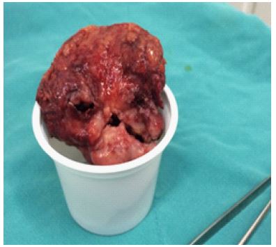
A 55-year-old, 165 cm tall, 70 kg male patient was referred to our lower right side, which started about three days ago and whose severity gradually increased. In the physical examination of the patient, tenderness was found in the right lower quadrant, and defense and rebound were positive. The patient’s temperature was normal. It was learned in his history that he had undergone appendectomy 11 years ago. In the laboratory examination, WBC: 11300 103/uL, neutrophil: 8800 103/uL andA 55-year-old, 165 cm tall, 70 kg male patient was referred to our lower right side, which started about three days ago and whose severity gradually increased. In the physical examination of the patient, tenderness was found in the right lower quadrant, and defense and rebound were positive. The patient’s temperature was normal. It was learned in his history that he had undergone appendectomy 11 years ago. In the laboratory examination, WBC: 11300 103/uL, neutrophil: 8800 103/uL and other tests were found to be normal. Then, Computed Tomography, which was performed by an external center by giving oral and intravenous contrast material and evaluated with the preliminary diagnosis of “Panniculitis”, was re-evaluated. In the computed tomography examination, the appendix could not be visualized in the right lower quadrant. In the old appendectomy site, an oval-shaped area of approximately 4 x 5 cm in size associated with the peritoneum, with fat density compatible with fat necrosis and surrounded by soft tissue rims, was detected. With these findings, a diagnosis of omental infarction was made and conservative treatment was initiated. Then, in the evaluation made the next day, it was observed that the white blood cell count: 8000 103/uL neutrophil: 5500 103/uL tended to regress, but the patient’s right lower quadrant pain continued, but the sensitivity was the same. Again, defense and rebound were positive. It was decided to continue the conservative treatment of the patient. In the evaluation made the next day, WBC: 12900 103/uL and Neutrophil: 11900 103/uL were found and it was decided to operate on the patient. During the exploration, an infarcted omentum was detected adhering to the lateral wall in the old appendectomy lodge, and the infarcted omentum was excised (Figure 1). Histopathological examination revealed hemorrhage and infarction foci in the omentum. In microscopic examination, infarcted foci were occupied by acute inflammatory cells. The patient, whose pathology was not detected after the operation, was discharged on the 3rd day with recovery.
Discussion and Conclusion
Omental infarction occurs as a result of torsion of the omentum around its long axial axis and compression of its vascular structure [1]. It may develop secondary to various causes such as vascular pathologies, hypercoagulable conditions and hernias, or it may develop idiopathic (primary) [2,3]. The causes of primary omental infarction are still unknown. Venous malformations that may cause stasis or thrombosis due to anatomical variation in vascular structures, and venous congestion developing after excessive and heavy meals are among the reasons suggested. It occurs twice as often in men than in women. It is also more common in obese individuals [3].
Pain is usually located in the right lower quadrant. Moderate leukocytosis, fever and vomiting may accompany the picture [4]. In our case, right lower quadrant pain accompanied the picture. However, we could not decide whether the torsion developed secondary to a previous appendectomy operation or it was most commonly located in the right lower quadrant as stated in the literature. The main point to be discussed here is; When omental infarct develops, should the patient be followed with conservative treatment methods or should resection be performed? Puylaert and Vriesman reported that no complications developed in the patients they treated with conservative treatment methods [4]. Therefore, there is no consensus on the treatment of patients with omental infarction. While some authors argue for the need for surgical treatment by suggesting possible complications, others argue that late complications are not as common as people think and conservative treatment methods will be sufficient [2,3,5]. In our case, we started to apply a conservative treatment method to the patient, but since we could not get any results, we provided the patient’s recovery with surgical treatment.
References
- McClure MJ, Khalili K, Sarrazin J, Hanbidge A. Radiological features of epiploic appendagitis and segmental omental infarction. Clin Radiol. 2001; 56: 819-27.
- Puylaert JB. Right-sided segmental infarction of the omentum: Clinical, US, and CT findings. Radiology 1992; 185: 169-72.
- Grattan-Smith JD, Blews DE, Brand T. Omental infarction in pediatric patients: Sonographic and CT findings. AJR Am J Roentgenol. 2002; 178: 1537-9.
- Puylaert JB. Right-sided segmental infarction of the omentum: Clinical, US, and CT findings. Radiology. 1992; 185: 169-72.
- Wiesner W, Kaplan V, Bongartz G. Omental infarction associated with right-sided heart failure. Eur Radiol. 2000; 10: 1130-2.