
Journal of Clinical Images and Medical Case Reports
ISSN 2766-7820
Case Report - Open Access, Volume 4
A case of Turner syndrome associated with juvenile idiopathic arthritis
Azadeh Zeinab Mirzaee1; Samin Afazel1; Shabnam Hajiani Ghotbabadi2; Reza Shiari3
1Department of Pediatrics, Masih Daneshvari Hospital, Shahid Beheshti University of Medical Sciences, Tehran-Iran.
2Department of Pediatrics, Shiraz University Hospital, Namazi Hospital, Shiraz-Iran.
3Department of Pediatrics, Mofid Children’s Hospital, Shahid Beheshti University of Medical Sciences, Tehran-Iran.
*Corresponding Author : Reza Shiari
Department of Pediatrics, Mofid Children’s Hospital, Shahid Beheshti University of Medical Sciences, Tehran-Iran.
Tel: +98-21-22227033;
Email: shiareza@yahoo.com
Received : Jul 05, 2023
Accepted : Jul 25, 2023
Published : Aug 01, 2023
Archived : www.jcimcr.org
Copyright : © Shiari R (2023).
Abstract
Turner’s syndrome is a chromosomal disorder characterized by complete or partial monosomy of the X chromosome. It is possible for Turner’s syndrome to be associated with autoimmune diseases such as rheumatoid arthritis. There is a limited number of reports in the literature about the association of Turner’s syndrome and Juvenile idiopathic arthritis. In this article we discuss about a patient with Turner’s syndrome who was presents with swelling of her left knee which was suggestive of oligo-Juvenile idiopathic arthritis.
Keywords: Turner syndrome; Juvenile idiopathic arthritis.
Citation: Mirzaee AZ, Afazel S, Ghotbabadi SH, Reza S. A case of Turner syndrome associated with juvenile idiopathic arthritis. J Clin Images Med Case Rep. 2023; 4(8): 2524.
Introduction
Juvenile Idiopathic Arthritis (JIA) is an autoimmune, inflammatory joint disease. It is the most common rheumatic disease in children and one of the more common chronic illnesses of childhood. JIA is an umbrella term for arthritis of unknown origin, lasting for more than 6 weeks with onset age of less than 16 years [1]. Oligoarthritis is defined as involvement equal and less than 4 joint within the first six months of disease onset predominantly affecting the large joint of lower extremities such as knee and ankles [2].
Turner’s Syndrome (TS) is a condition characterized by complete or partial monosomy of the X chromosome and defined by combination of phenotypic features. Half of the patients with Turner’s syndrome have a 45X hromosome complement. Turner syndrome occurs in approximately 1 in 2000 to 1 in 2500 live female births. Clinical findings in the newborns can include small size for gestational age, webbing of the neck, protruding ears, and lymphedema of the hands and feet, although many newborns are phenotypically normal [3].
Recent studies have suggested that there is a higher incidence of Autoimmune Diseases (AD) in people with Turner syndrome [4].
In this paper, we report a 4-year-old Iranian girl with Turner syndrome who was complicated with JIA.
Case presentation
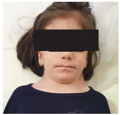
The patient is a 4-year-old Iranian girl born after an uneventful pregnancy and normal delivery. She was the third child of non-consanguineous, phenotypically normal parents. Her older siblings are healthy without any medical condition. At the age of 2 years dysmorphic features were noticed, including bilateral blepharoptosis, long philtrum, low set ears, webbing of the neck (Figure 1). She also had failure to thrive, ambiguous genitalia and developmental delay. Her cousin on the father side had FTT and ambiguous genitalia and died at the age of 6 month.
She was diagnosed with Turner syndrome via genetic study which revealed mixed gonadal dysplasia compatible with 45XO/46XY (Figure 2), compatible with abnormal chromosomal complements with mosaicism. Therefor she had undergone bilateral gonadectomy (oophorectomy) and lobectomy via laparoscopic surgery. In the biopsy there were mixed ovarian and testicular tissue in gonads and fallopian tube and epididymal tissue in the presumed fallopian tube.
At the age of 4 years, she was referred to Mofid Children Hospital complaining of progressive left knee swelling and pain for the past two months and inability to walk. She didn’t have any history of trauma to the affected knee. She didn’t have any episodes of fever.
In physical examination, the appearance of left knee was abnormal with swelling, tenderness and mild warmness. The Ballottement test of the left knee was positive and she also had reduced range of motion on it. There was no other joints involvement. She didn’t have any skin rashes. She was underweighting (<3th PC) and short statured (<3th PC).
The ultrasound of left knee showed mild effusion about 6 mm with no synovial thickening (Figure 3). With suspicion of septic arthritis she underwent arthrocentesis that was negative for septic arthritis.
In lab data, CBC was normal (WBC: 9600/μl, Hb: 10 g/dl, PLT: 397000/μl), acute phase reactants were high (ESR: 35 mmHg, CRP: 52 mg/dl) and Rheumatoid factor, Wright, 2ME and PPD were all negative. Blood culture and urine culture showed no bacterial growth after 24 hours. Biochemistry lab tests were normal (BUN: 7.3 mg/dl, Cr: 0.6 mg/dl, AST: 25 U/L, ALT: 11 U/L, uric acid: 2 mg/dl, LDH: 416 U/L). Analysis of the synovial fluid was follows: (Glu: 35 mg/dl, Pro: 3100 gr/dl, WBC: 6000/mm3, PMN: 80%, MN: 20%, RBC: 80/mm3, LDH: 2201 U/L). The synovial fluid smear and culture were was also negative. According to her lab results septic arthritis, septicemia, Tuberculosis and Brucellosis were all ruled out. the patient fulfilled the International League of Associations for Rheumatology (ILAR) criteria for a diagnosis of oligoarticular JIA [2]. Her ophthalmology examination was normal and there were no evidence of uveitis or iridocyclitis.
She was treated with intra-articular injection of Triamcinolon and oral Naproxen.
The patient was also referred to Endocrinologist because of poor weight gain and short stature.


Discussion
We have described a 4-year-old Iranian girl with Turner syndrome who was referred because of pain and swelling in her left knee. The diagnosis of rheumatoid disease in our patients has been a subject of debate, as Juvenile Idiopathic Arthritis (JIA) is an exclusion diagnosis and therefore all the other differential diagnosis has to be ruled out including septic arthritis which is the most important one as it could have long term and irreversible complications such as leg-length discrepancy, abnormalities of bone growth, limitation of movement and osteomyelitis [5]. Other conditions which could cause the patient’s symptoms include Septicemia, Tuberculosis and Brucellosis [6]. In the case of our patient all of these conditions were ruled out via laboratory findings and clinical tests.
The prevalence of JIA in patients with TS seems to be at least six times greater than would be expected if the two conditions were only randomly associated [7]. There is a theory which suggests that the abnormalities of the X chromosome may influence immune tolerance, making TS patients more susceptible to autoimmune diseases [8]. The underlying immunopathogenic mechanism of this association remains partially unexplained. Studies have suggested a higher responsibility of the X chromosome abnormalities. For instance, the long arm of the X chromosome hosts a MHC-locus, so the loss of that region in Turner syndrome may cause a deficiency in immune regulation [9].
In recent studies, it has been found that there is a higher incidence of Autoimmune Diseases (AD) in the population with TS. According to the latest guidelines the risk of AD increases with age; therefore, it has been recommended that both children and adults with TS, do regular follow-ups and screenings.
References
- Martini A, Lovell DJ, Albani S, Brunner HI, Hyrich KL, et al. Juvenile idiopathic arthritis. Nature Reviews Disease Primers. 2022; 8: 5.
- Petty RE, Southwood TR, Manners P, Baum J, Glass DN, et al. International League of Associations for Rheumatology classification of juvenile idiopathic arthritis: second revision, Edmonton, 2001. The Journal of rheumatology. 2004; 31: 390-2.
- Kikkeri NS, Nagalli S. Turner Syndrome. StatPearls: StatPearls Publishing. 2021.
- Gawlik AM, Berdej-Szczot E, Blat D, Klekotka R, Gawlik T, et al. Immunological profile and predisposition to autoimmunity in girls with Turner syndrome. Frontiers in Endocrinology. 2018; 9: 307.
- Akinyoola A, Obiajunwa P, Oginni L. Septic arthritis in children. West African journal of medicine. 2006; 25: 119-23.
- Giancane G, Consolaro A, Lanni S, Davi S, Schiappapietra B, et al. Juvenile idiopathic arthritis: diagnosis and treatment. Rheumatology and therapy. 2016; 3: 187-207.
- Zulian F, Schumacher H, Calore A, Goldsmith D, Athreya B. Juvenile arthritis in Turner’s syndrome: A multicenter study. Clinical and experimental rheumatology. 1998; 16: 489-94.
- Wihlborg CEM, Babyn PS, Schneider R. The association between Turner’s syndrome and juvenile rheumatoid arthritis. Pediatric Radiology. 1999; 29: 676-81.
- Lleo A, Moroni L, Caliari L, Invernizzi P. Autoimmunity and Turner’s syndrome. Autoimmunity reviews. 2012; 11: A538-A43.