Journal of Clinical Images and Medical Case Reports
ISSN 2766-7820
Clinical Image - Open Access, Volume 4
Unusual presentation of Burkitt lymphoma with late onset secondaries
Praveen Sandeep1; Gopinathan2; Aarthi Deepesh3; Milly Mathew4; Georgi Abraham5*
1Registrar, Department of Nephrology, MGM HealthCare, Chennai, India.
2Department of Hematologist, MGM HealthCare, Chennai, India.
3Department of Neuroimaging and Interventional Radiology, MGM HealthCare, Chennai, India.
4Senior Consultant, Department of Nephrologist, MGM HealthCare, Chennai, India.
5Department of Nephrologist, MGM HealthCare, Chennai, India.
*Corresponding Author : Georgi Abraham
Department of Nephrologist, MGM HealthCare, Chennai, India.
Email: abraham_georgi@yahoo.com
Received : May 19, 2023
Accepted : Aug 17, 2023
Published : Aug 24, 2023
Archived : www.jcimcr.org
Copyright : © Abraham G (2023).
Citation: Sandeep P, Gopinathan, Deepesh A, Mathew M, Abraham G. Unusual presentation of Burkitt lymphoma with late onset secondaries. J Clin Images Med Case Rep. 2023; 4(8): 2559.
Clinial image description
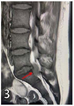
The diagnosis of endemic Burkitt lymphoma was made in this 16 year old Sudanese boy by kidney biopsy histopathology (Figure 1) and touch imprint (Figure 2) as the bone and bone marrow biopsies were unremarkable. He was given 7 cycles of R-COPADM1 chemotherapy according to LMB-89 protocol, and he became disease free by PET scan. Subsequently, he presented 3 months later with severe lower back ache and a cauda equina lesion. Figure 3 shows L4-L5 disc prolapse and secondary deposits (red arrow) in the spinal canal. Surgical excision of the disc and the spinal canal lesion done.
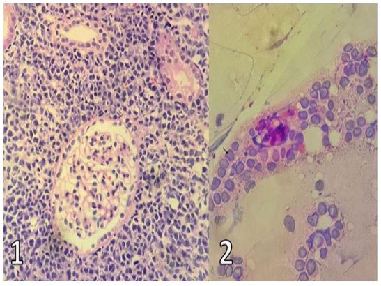
Figure 2: Touch Imprint of kidney biopsy.