
Journal of Clinical Images and Medical Case Reports
ISSN 2766-7820
Clinical Image - Open Access, Volume 4
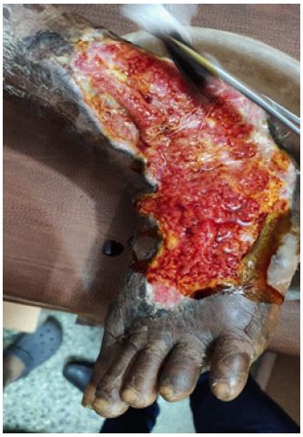
Severe pyoderma gangrenosum: A rare clinical image
Ashwin Gokhare*; Shweta Parwe*
Department of Panchakarma, Mahatma Gandhi Ayurved College Hospital and Research Centre, Datta Meghe Institute of Higher Education & Research, Sawangi, Wardha, Maharashtra, India.
*Corresponding Author : Ashwin Gokhare & Shweta Parwe
Department of Panchakarma, Mahatma Gandhi Ayurved College Hospital and Research Centre, Datta Meghe Institute of Higher Education & Research, Sawangi, Wardha, Maharashtra, India.
Email: gokhareashwin@gmail.com &
Shweta.parwe@dmimsu.edu.in
Received : Aug 02, 2023
Accepted : Aug 22, 2023
Published : Aug 29, 2023
Archived : www.jcimcr.org
Copyright : © Gokhare A, Parwe S (2023).
Keywords: Pyoderma gangrenosum; Rheumatoid disease; Haematological disease; Neutrophilic dermatosis.
Citation: Gokhare A, Parwe S. Severe pyoderma gangrenosum: A rare clinical image. J Clin Images Med Case Rep. 2023; 4(8): 2565.
Description
Pyoderma Gangrenosum (PG) is a very uncommon neutrophilic dermatosis that is not contagious. According to the clinical definition, it begins clinically as sterile pustules that quickly advance into painful ulcers of varying depth and size with weakened violaceous margins. The most frequently impacted area is the leg, however, other skin and mucous membrane areas may also be affected. The exact etiology has not yet been established [1,2]. The course may be chronic or relapsing, moderate, malignant, or extremely morbid. In a lot of instances, PG is linked to an underlying illness, most frequently inflammatory bowel disease, rheumatoid or hematological disease, or cancer.PG occurs most commonly on the lower legs with a preference for the pretibial area. On the other hand, less than 3% of patients with Crohn’s disease or ulcerative colitis develop PG [3]. The history of an underlying condition, the usual clinical maniKeywords festation, the histology, and the elimination of other conditions that might manifest similarly are all used to make the diagnosis of PG. Women are more frequently affected than men and incidence peaks between the ages of 20 and 50.
References
- Van den Driesch P. Pyoderma gangrenosum: A report of 44 cases with follow-up. Br J Dermatol. 1997, 137: 1000-1005.
- Graham JA, Hansen KK, Rabinowitz LG. Pyoderma gangrenosum in infants and children. Pediatr Dermatol. 1994, 11: 10-17.
- Ozdil S, Demir K, Boztas G. Crohn’s disease: Analysis of 105 patients. Hepatogastroenterology. 2003; 50: cclxxxvii-ccxci.