Journal of Clinical Images and Medical Case Reports
ISSN 2766-7820
Clinical Image - Open Access, Volume 4
Calcified coronary artery aneurysm
Kaavya Nair1*; Nandan Anavekar2
1Research Fellow, Mayo Clinic, Rochester MN, USA.
2Program Director for Adult Cardiovascular Medicine, Mayo Clinic, USA.
*Corresponding Author : Kaavya Nair
Research Fellow, Mayo Clinic, Rochester MN, USA.
Phone: 818-451-5294;
Email: kaavyanjali96@gmail.com
Received : Aug 03, 2023
Accepted : Aug 23, 2023
Published : Aug 30, 2023
Archived : www.jcimcr.org
Copyright : © Nair K (2023).
Keywords: Cardiology; Imaging; Decision-making.
Citation: Nair K, Anavekar N. Calcified coronary artery aneurysm. J Clin Images Med Case Rep. 2023; 4(8): 2567.
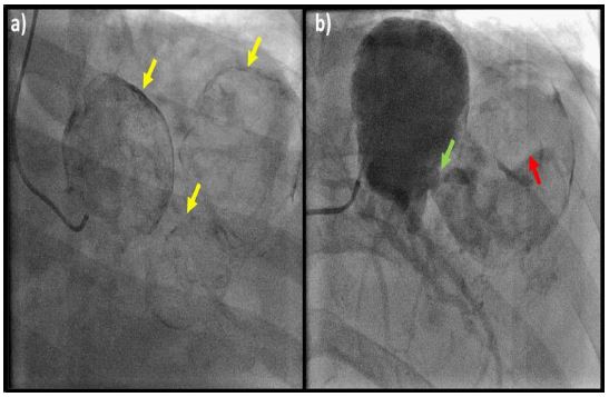
Description
58-year-old woman presented to the hospital with sudden onset right sided abdominal and chest pain. Coronary angiogram demonstrated calcified aneurysms (yellow arrows) at the bifurcation left main which were in communication with one another (green arrow). The distal aneurysm was noted to be partially thrombosed (red arrow) (Figure 1).
Cardiothoracic surgery did not recommend any surgery to coronary arteries, given stability of aneurysm size when compared to prior imaging. Notably, the patient was found to have a large pericardial effusion without signs of tamponade. After pericardiocentesis, there was a marked improvement of symptoms.
Funding: None
Conflicts of interest: None.