
Journal of Clinical Images and Medical Case Reports
ISSN 2766-7820
Case Report - Open Access, Volume 4
Successful drug challenge to a different drug formulation in fixed drug eruption
*Corresponding Author : Ali Alzahrani
Division of Molecular Endocrinology, Department of Molecular Oncology, King Faisal Specialist Hospital and Research Centre, P.O.Box 3354, Research Center (MBC 03), Riyadh, 11211, Saudi Arabia.
Email: alithesefi@hotmail.com
Received : Aug 07, 2023
Accepted : Aug 28, 2023
Published : Sep 04, 2023
Archived : www.jcimcr.org
Copyright : © Alzahrani A (2023).
Abstract
Type 4 hypersensitivity reactions convey a number of conditions that include Fixed drug eruptions. They share similar pathophysiologic backgrounds and sometimes presentation but can have very variable prognostications. Drugs are amongst the possible causes with acetaminophen and other NSAIDs being reported very frequently. We present a case of a patient reacting to flavoured oral ibuprofen and acetaminophen formulations exhibiting fixed drug eruptions with bullae formation. We describe our successful challenge to non-flavoured acetaminophen and ibuprofen. We briefly discuss fixed drug eruptions in regard to their incidence, pathophysiology and management. Introduction Fixed drug eruptions are skin reactions that results from a Type 4 hypersensitivity reaction potentially developing within minutes to hours or occasionally more akin to other type 4 reactions but may happen 14 days after an offending agent (for example drug or vaccine) [1]. In fixed drug eruptions, the lesions have this tendency to reappear on the same places upon further exposure hence the term “fixed “ and at sites of previous trauma [2]. This phenomenon can be helpful in diagnosing and predicting the distribution of the reaction. It is classically described as usually being oval with a dusky colour and being well demarcated. These lesions, however, can erupt variably and express drastically different features similar to Erythema Multiforme or more dramatically bullous eruptions that mimic other type 4 reactions known for worse prognosis like Toxic Epidermal Necrolysis and Steven Johnson Syndrome. Lack of mucosal involvement and the rash distribution can give diagnostic hints [3] but not conclusive as mucosal involvement has been described [4]. Systemic symptoms can occur at a rarity, but they are more common in bullous eruptions. Diagnosis remains largely clinically though sometimes; biopsy is needed to differentiate, and prognosis is general very favourable compared to TEN/SJS. A case control study revealed otherwise mainly in older age [3,5]. Many drugs have been described and reported including the very commonly used NSAIDs and acetaminophen [1,6]. We couldn’t find enough data highlighting the occurrence of over-the-counter medications FDE, but it has been reported to variable OTC drugs including multivitamins [7]. We came across a case of an 11-year-old girl that demonstrated fixed drug eruptions to flavoured liquid formations of NSAIDs and Tylenol, but not to plain formulations of both. To our knowledge this is the first case report demonstrating the occurrence of drug eruptions of a NSAIDs/acetaminophen that successfully tolerated non-flavoured formulations upon drug challenge.
Citation: Alzahrani A. Successful drug challenge to a different drug formulation in fixed drug eruption. J Clin Images Med Case Rep. 2023; 4(9): 2574.
Case presentation
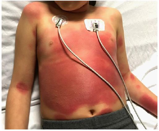
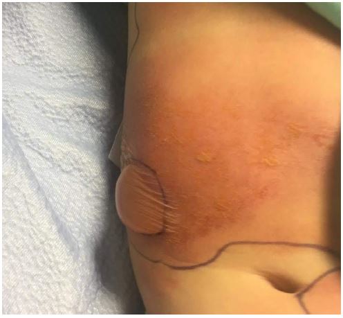
An 11-year-old female who has a history of previous Fixed Drug Eruption in 2019 with suggestive biopsy finding to consuming flavoured Ibuprofen. She had bulla formations and rash that spread over her neck and chest that was managed with opioids, antihistamines and topical steroids. She was scheduled for an acetaminophen challenge, but the family was reluctant. 2 years later, she had an ER visit for a viral. She was given non flavoured liquid acetaminophen. She had no reaction and was discharged on non-flavoured acetaminophen to no issues at home. A month after, she went camping and a few days upon return, she started having fever and rhinorrhoea. Swab for COVID-19 was negative and the patient was given flavoured acetaminophen liquid that contained anhydrous citric acid, FD&C red no. 40 (Allura red, a dye), glycerin, high fructose corn syrup, microcrystalline cellulose, carboxymethylcellulose sodium, purified water, sodium benzoate, sorbitol solution, sucralose and xanthan gum. Within 3 hours, the patient started to have burning sensation on the sites she previously had a rash 2 years ago that included her neck, chest and abdomen where she also started to have increasing pain. The rash composed of multiple erythematous plaques of various sizes with vesicular/bullous components as shown (Figure 1). She was vitally stable. She was admitted for monitoring. She had normal ECG at that point, her labs demonstrated normal complete blood count, normal liver function test and normal kidney function. Cetirizine and topical steroids were started. Within the two days after she developed a bulla on the right side of her abdomen (Figure 2). The bulla wasn’t tense but slightly painful. Dermatology thought a biopsy wasn’t needed given the lesions followed expected distribution of her previous fixed drug eruption. The patient continued to have no mucosal or genital involvement. The bullae continued to fill (Figure 3) before it ruptured on day 6 and the patient was discharged home on topical steroids. Complete resolution of active lesions took 3 weeks. She is left with hyperpigmentation on her abdomen and neck.

Lymphocyte toxicity assay, an in vitro blood test was done by clinical pharmacy. The patient’s lymphocytes isolated from peripheral blood samples showed variable hypersensitivity results to flavoured oral formulations of acetaminophen and ibuprofen. Non flavoured formulation showed no reaction. We have offered them a patch test to both flavoured brands she reacted with in addition non flavoured ones. We thought this was the safest plan given her reactions were more generalized and necessitated admission. Unfortunately, due to the acuity of their schedule, they declined. We discussed with them the safety and concerns of doing a challenge. Given the lymphocyte toxicity assay showed complete lack of reaction to non-flavoured formulations of both medications, we decided to do a challenge to both medications. We started with the non-flavoured liquid ibuprofen challenge first and we aimed for a total dose of 100 mg. We decided to start with one tenth of a dose starting at 10 mg and watched clinically for 3 hours. Vitals were taken at the start. She was frequently inspected for any skin rashes. She had no issue and given the remaining dose and kept for 3 more hours with frequent inspection. At the end, she reported no reaction to the challenge and was completely normal. We have phone called her twice over two weeks to no issues. She came to clinic again for one step non flavoured liquid acetaminophen challenge that she had no reaction to. She reported no issues upon virtual follow ups. We advised her to take non flavoured formulations of the over-the-counter medications as of now and to reconsider patch testing to the flavoured formulations when that’s convenient. Our aim is to try an elicit possible ingredients other than the active medications causing the reaction.
Discussion
Fixed drug eruptions remain a rare medical entity that composed around 0.003% of total skin reactions in a US based study that included 2.7 million individuals [8]. It is however reported more frequently in smaller series studies. Two studies showed a 14.1% over a series of 90 patients and as done in Tunisia [9] and 22% over 50 patients in a study conducted in India [10]. While drugs are the most common cause, other culprits such as vaccines and food have been prescribed [1]. They also tend to occur at sites of previous trauma or injury suggesting a recall phenomenon [2]. They usually have a good prognosis, but not always and can lead to local complication as well such as eyelid necrosis [11]. The pathophysiology of fixed drug eruptions is majorly driven by intraepidermal CD8+ cells found in active and inactive lesions [12]. These cells upon reactivation with a causative agent, trigger interferon gamma in addition to cytotoxic granules and granzyme b contributing to the development of the skin lesions. Histopathology for fixed drug eruptions differ depending in the stage and occasionally can be inconclusive. If the biopsy was taken at an active lesion, it shows basal keratinocytes degradation and lymphocytic infiltration in the dermis. Biopsy of resting lesions show numerous CD8 lymphocytes in the dermo-epidermal junction that moves upward while maintaining a normal looking dermis 2-3 hours upon being triggered then after 48 hours, the active features appear. There are other atypical presentation including leukocytoclastic vasculitis [13]. It has been shown in studies that primed CD8 cells can survive in the absence of a trigger for more than 4 years further explaining the repeated involvement of same skin sites upon reactivation by a trigger [14]. While acetaminophen and NSAIDs are thought to be one of the most common triggers for the variable forms of fixed drug eruptions [1,6]. However, other culprits including antibiotics and anti-epileptics are also reported with variable frequency. Dyes and colouring agents were implicated in many reactions including both immediate and delayed [15,16], but no specific case reports with successful challenges. There has been variable reports about the reactions ranging from the typical presentation to other unique but individual presentations such as the aforementioned eyelid necrosis [11]. Diagnosing fixed drug eruption remains largely clinical thanks to the typical presentation and the usually fixed re-involvement of previously affected areas. A biopsy is helpful but isn’t always indicated especially in the complete lack of worrisome or atypical features or there are highly suspicions of TEN or SJS. Lymphocyte toxicity assay is a novel diagnostic test that has been adapted to variable extents clinically to help identify a causative agent for delayed drug reactions [17] The idea of this in vitro test is to see how the sensitive lymphocyte of an individual is unable to detoxify the susceptible drug applied in vitro. It has garnered some evidence but for the most part has remained largely a supportive test as in studies it has demonstrated variable sensitivity and specificity [18]. It remains not largely available and uncommon [17]. Multiple case reports have demonstrated it’s use [19,20]. It doesn’t substitute clinical history, patch testing or oral challenge if feasible to diagnose type 4 reactions. In our case, it was done beforehand by clinical pharmacy when they were consulted. While the results were consistent with our clinical history, it was the fact that she tolerated the non-flavoured acetaminophen given to her in an ER visit that prompted us to offer the oral challenge. Fixed bullous drug eruption treatment follows the principals of type 4 reactions where the first step is to stop all agents implied and then to treat symptomatically. Care should be given to differentiate between the different type 4 reactions. Itching can be managed with H1 anti-histamines and topical variable potency steroids. In case oral involvement, local analgesics such as lidocaine can be utilized. In case of pain, appropriate analgesia should be given. Systemic immunosuppressants such as Cyclosporine are indicated sometimes in case of generalized disease [17] Provocation challenges are advisable if the culprit drug can’t be identified clinically in cases where more are suspected. It’s however contraindicated in the presence of severe systemic symptoms or if SJS and TEN were highly suspected and not ruled out. No way has been standardized however and the practice depend largely on the physician experience. A recommended way is to start with a 1/10th then increase the dose to a full dose every 12 to 24 hours. Another approach is to give half a dose then increase it to full dose if no reaction occurs. In our approach, we managed to a 1/10th of a dose with remaining dose given in 3 hours while watching her for 3 more hours. Another approach we considered was to space the challenge over 2 days course, but the patient refused given time constraint. With our non-flavoured acetaminophen, we have administered it in one dose and followed her half a clinic day (4 hours) as she has been given that before to no reaction and our challenge was more to affirm that. Patch testing remains largely non-standardized for the most part and follows different protocols. The reactions would be localized. We wanted to mix the flavoured liquid medications with petrolatum and have it occluded on her skin if erythema developed within 24 hours and lasted more than 6 hours, the test is positive. This could have helped prove reactivity and usability of the patch test to her known two medications after which point, we could have added the inactive ingredients that are available with other over the counter medications and see if she will have positives as well. That will help us avoid further possible reactions and give us clues about the possible inactive ingredient/component leading to her reaction. Of note, the test is limited by high degree of false negative especially when applied to non-involved area reaching to 40% [21] but remains a very useful test if used in the right clinical setting.
Conclusion
Fixed drug eruptions are one the type 4 hypersensitivity reactions. It can present variably and be mistaken of other drug reactions or primary skin conditions. It usually has a good clinical outcome. NSAIDs and acetaminophen remain among the most common causes of fixed drug eruptions, but as in our case, we wonder if those reactions are sometimes caused by components of different formulations of these medications other than the active ingredient. Considering that with appropriate and delicate clinical assessment using patch testing and oral challenge, if possible, would help keep these otherwise safe, affordable, and commonly needed medications as an available option.
References
- McClatchy J, Yap T, Nirenberg A, et al. Fixed drug eruptions - the common and novel culprits since 2000. J Dtsch Dermatol Ges.2022; 20: 1289-1302.
- Mizukawa Y, Shiohara T: Trauma-localized fixed drug eruption: Involvement of burn scars, insect bites and venipuncture sites. Dermatology. 2002; 205: 159-61.
- Cho YT, Lin JW, Chen YC, et al. Generalized bullous fixed drug eruption is distinct from Stevens-Johnson syndrome/toxic epidermal necrolysis by immunohistopathological features. J Am Acad Dermatol. 2014; 70: 539-48.
- Ferreira C, Corrales T, Guilherme A. Fixed Drug Eruption on the Tongue Due to Naproxen: J Investig Allergol Clin Immunol. 2020; 358-359.
- Lipowicz S, Sekula P, Ingen-Housz-Oro S, et al. Prognosis of generalized bullous fixed drug eruption: Comparison with Stevens-Johnson syndrome and toxic epidermal necrolysis. Br J Dermatol. 2013; 168: 726-32.
- Brahimi N, Routier E, Raison-Peyron N, et al. A three-year-analysis of fixed drug eruptions in hospital settings in France. Eur J Dermatol. 2010; 20: 461-4.
- Jha N. Bullous fixed drug eruption related to multivitamins. Dermatol Online J. 2020; 15: 13030-3150.
- Wong A, Seger DL, Lai KH, et al. Drug Hypersensitivity Reactions Documented in Electronic Health Records within a Large Health System. J Allergy Clin Immunol Pract. 2019; 7: 1253-1260.
- Khaled A, Kharfi M, Ben Hamida, et al. Cutaneous adverse drug reactions in children. A series of 90 cases. Tunis Med. 2012; 90: 45-50.
- Sharma VK, Dhar S. Clinical pattern of cutaneous drug eruption among children and adolescents in north India. Pediatr Dermatol. 1995; 12: 178-83.
- Kimmatkaar P, Das S, Gandhi A, et al. Paracetamol-induced fixed drug eruption presenting as eyelid skin necrosis. Indian J Ophthalmol. 2018; 66: 1627-1629.
- Mizukawa Y, Shiohara T. Fixed drug eruption: A prototypic disorder mediated by effector memory T cells. Curr Allergy Asthma Rep. 2009; 9: 71-7.
- Harris A, Burge SM. Vasculitis in a fixed drug eruption due to paracetamol. Br J Dermatol. 1995; 133: 790-1.
- Mizukawa Y, Yamazaki Y, Shiohara T. In vivo dynamics of intraepidermal CD8+ T cells and CD4+ T cells during the evolution of fixed drug eruption. Br J Dermatol. 2008; 158: 1230-8.
- Tattersall I, Reddy BY. Fixed Drug Eruption due to Achiote Dye. Case Rep Dermatol. 2016; 28: 14-8.
- Caballero ML, Quirce S. Delayed Hypersensitivity Reactions Caused by Drug Excipients: A Literature Review. J Investig Allergol Clin Immunol. 2020; 400-408.
- Patel S, John, et al. Fixed Drug Eruptions: An Update, Emphasizing the Potentially Lethal Generalized Bullous Fixed Drug Eruption. Am J Clin Dermatol. 21: 393-399.
- Elzagallaai AA, Jahedmotlagh Z, Del Pozzo-Magaña BR, et al. Predictive value of the lymphocyte toxicity assay in the diagnosis of drug hypersensitivity syndrome. Mol Diagn Ther. 2010; 14: 317-22.
- Kim MH, Shim EJ, Jung JW, Sohn SW, Kang HR. A case of allopurinol-induced fixed drug eruption confirmed with a lymphocyte transformation test. Allergy Asthma Immunol Res. 2012; 4: 309-10.
- Demir S, Cetin EA, Unal D, et al. Generalized Fixed Drug Eruption Induced by Fluconazole Without Cross-Reactivity to Itraconazole: Lymphocyte Transformation Test Confirms the Diagnosis. Drug Saf Case Rep. 20182; 5: 2.
- Phillips EJ, Bigliardi P, Bircher AJ, Shear NH, Tanno LK, et al. Controversies in drug allergy: Testing for delayed reactions. J Allergy Clin Immunol. 2019; 143: 66-73.