
Journal of Clinical Images and Medical Case Reports
ISSN 2766-7820
Case Report - Open Access, Volume 4
Hemangioma of the petrous apex: Symptoms and diagnosis approach
L Douimi*; C Rsaissi; Y Oukessou; S Rouadi; R Abada; M Roubal; M Mahtar
ENT Head and Neck Surgery Department, Hospital IBN ROCHD University Hospital, Faculty of Medicine and Pharmacy, Hassan II University, Casablanca, Morocco.
*Corresponding Author : L Douimi
ENT Head and Neck Surgery Department, Hospital IBN ROCHD University Hospital, Faculty of Medicine and Pharmacy, Hassan II University, Casablanca, Morocco.
Email: loubnadouimi@gmail.com
Received : Aug 15, 2023
Accepted : Sep 11, 2023
Published : Sep 18, 2023
Archived : www.jcimcr.org
Copyright : © L Douimi (2023).
Abstract
This study reports a rare case of a large petrous apex hemangioma. A 17-year-old boy presented with hemifacial spasm and dysphonia, followed by right oculomotor dysfunction, ataxia, and right tongue deviation. Initial CT-SCAN showed massive heterogeneous tissue process located in the petrous apex, enhanced after contrast injection with significant osteolysis. Brain MRI showed a large oval formation centered on the right petrous bone, intermediate T1 signal, heterogeneous T2 hyperintense, focal and heterogeneous amplification after gadolinium injection. Biopsies were performed under general anesthesia, revealing morphological aspects and immunophenotypes favorable to capillary hemangiomas.
Temporal bone hemangiomas can mimic other, more common basal tumors. Imaging usually does not provide a definitive preoperative diagnosis, however surgical resection and a biopsy can.
Keywords: Capillary hemangioma; Petrous apex; Temporal bone; Diagnosis; Imaging aspect.
Citation: Douimi L, Rsaissi C, Oukessou Y, Rouadi S, Abada R, et al. Hemangioma of the petrous apex: Symptoms and diagnosis approach. J Clin Images Med Case Rep. 2023; 4(9): 2595.
Introduction
Hemangiomas are rare benign vascular tumors. They are more common in the head and neck region and can be classified as capillary, cavernous, or mixed. They are usually found in infancy on the skin or mucous membrane surfaces of the mouth and nose. Children usually have onset by 1 year of age and resolve at the age of 5 or 6 years old [1]. Capillary hemangiomas are capillary-like ducts found in the skin, subcutaneous tissue, lips, liver, spleen, and kidneys [2].
There has been some misunderstanding in the literature regarding the terminology of vascular lesions, the term hemangioma has been misused to describe what appear to be vascular malformations [3,4], Although vascular lesions are common in the head and neck, [5] their localization in the temporal bone are rare, they are usually located in the internal auditory canal or along the facial nerve, due to the abundant blood supply surrounding the Scarpa and geniculate ganglion [6]. Few cases of middle and outer ear localization have been documented [7].
We report the case of a 17-year-old boy with osteolytic hemangioma in the right petrous bone associated with a hemifacial spasm, a dysphonia, an ataxia and right oculo-motor disorders.
The purpose of this report is to discuss diagnostic methods. The characteristics of this disorder are also discussed on the basis of a review of the relevant literature.
Case presentation
A 17-year-old boy presented with a year and a half history of hemifacial spasm and dysphonia, followed by right oculo-motor disorders, an ataxia and right deviation of the tongue.
Clinical examination revealed a facial paralysis grade II with sign of Souques (paralysis of the VII nerve: Figure 1a), paralysis of the right vocal cord (nerve X), damage of the nerve VI (Figure 1b), IX/XI (curtain sign) and XII (Figure 1c).
Otoscopic examination showed a whitish, fleshy mass occupying the entire external auditory canal (Figure 2).
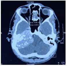
The initial CT-SCAN demonstrated voluminous heterodense tissue process of the petrous apex enhancing after injection of contrast product with important bone lysis, invading the internal auditory canal, the internal ear and a partial lysis of the walls of the right carotid canal pushing back the cerebral trunk and the 4th ventricle (Figure 3).
Cerebral MRI showed a coarse oval formation centered on the right petrous bone, well limited, with irregular contours, intermediate T1 signal, heterogeneous T2 hypersignal, flair and diffusion, and seat of void of signals, intensely and heterogeneously enhanced after injection of Gadolinium, realizing the aspect of salt and pepper. It measures 60.5 x 35 mm, and extends to 51 mm (Figure 4).
The various diagnoses considered were glioma, chondrosarcoma, tympano-jugular paraganglioma.
A biopsy under general anesthesia was performed and showed a morphological aspect and immunophenotype in favor of a capillary hemangioma.

Figure 1b: Oculo-motor disorder: Damage of the nerve VI.
Figure 1c: Deviation of the tongue: The XII nerve.
Figure 1: Clinical Symptoms of our case: Damage of the nerve VII (a), VI (b) , XII (c)


Discussion
There are few case reports of middle ear hemangioma; Osseous hemangiomas are reported as rare and usually affect the bone Spine, skull, but rarely temporal bone [8]. They occur mainly in areas rich in blood vessels (geniculate ganglion, inner ear canal) and depending on size or location they may cause facial paralysis, hemifacial spasm, hearing loss, tinnitus, otorrhea, ear pain, acute otitis media, or be asymptomatic [9,10]. In our case, the hemangioma seemed to be so advanced while the patient had many symptoms including hemifacial spasm, a dysphonia, right oculo-motor disorders, an ataxia and right deviation of the tongue.
These vascular lesions may be discovered incidentally during otoscopy by finding a mass in the EAC or in the presence of hearing impairment [11], such as the patients with conductive hearing loss.
In our case the otoscopy showed a whitish, fleshy mass occupying the entire external auditory canal.
Salamah Marzouqi and Halawani Roa reported to the otoscopy of the right ear a smooth, oval, well-circumscribed, reddish, pulsating, nontender mass approximately 1 cm in diameter, occupying two-thirds of the EAC. It is soft and compressible and appears to originate in the upper right posterior portion of the bony canal wall [12].
Once a lesion has been identified, it is important to determine its etiology because although glomus tumors are the most common vascular tumors of the middle ear, other diagnoses are possible: or inflammation (eosinophilic granuloma or cholesterol granuloma tumor) (rhabdomyosarcoma) [11,13]
If a hemangioma is suspected, High-Resolution Computed Tomography (HRCT) of the temporal bone, MRI, and angiography should also be performed. Computed Tomography (CT) of the temporal bone is the first choice to assess lesion location and size and middle ear involvement. Hemangiomas appear to have the same density as the brain parenchyma on CT scans and may develop ossification [14]. They appear as well-demarcated masses with limited contrast enhancement that are homogeneous during the proliferative phase and heterogeneous during the involutional phase [13].
Salamah Marzouqi and Halawani Roa reported a case with High-Resolution Contrast-Enhanced Computed Tomographic scanning (HR CECT) of the temporal bone showed a well-defined, rounded, homogenously enhancing lesion at the posterior-superior aspect of the right external auditory and whom the Magnetic Resonance Imaging (MRI) of the temporal bone showed a well-defined elongated right EAC lesion measuring 1.2 × 0.6 cm; the lesion appeared homogenous hyperintense on T2, hypointense on T1, and with homogenous enhancement on postcontrast sequence. There were vessels crossing anteriorly to the lesion [12].
Unlike our case whom the initial CT-SCAN demonstrated voluminous heterodense tissue process of the petrous apex enhancing after injection of contrast product and The cerebral MRI showed a coarse oval formation centered on the right petrous bone, well limited, with irregular contours, intermediate T1 signal, heterogeneous T2 hypersignal, Flair and diffusion, seat of void of signals, intensely and heterogeneously enhanced after injection of Gadolinium.
Preoperative HRCT is the preferred radiological evaluation method because it can reveal the size of the tumor, the presence of bone erosions, and the possibility of involvement of the ossicular chain [15]. To rule out the presence of a high jugular bulb or an abnormal carotid artery, careful review of radiographic findings is essential [16].
Because of their destructive and hemorrhagic nature, treatment of these tumors is essential. management based on complete tumor resection; indeed, the relatively high recurrence rate (16% to 23.3% in sinus locations) is directly dependent on the quality of surgical resection [13].
Many cases of spontaneous resolution have been reported [17]. Surgical technique depends on tumor onset, degree of hearing loss, and condition of the jugular bulb [13,18,19].
CO2 laser can be an interesting alternative to conventional surgery; it allows better visualization of middle ear structures by reducing bleeding [17,16,20].
Conclusion
Hemangiomas in the EAC and/or tympanic membrane are rare but must be considered in the differential diagnosis of EAC and/or tympanic membrane lesions. We report a case of right apex petrous hemangioma in a young boy which was discovered during otoscopy, in front of the appearance of a hemifacial spasm and a dysphonia then the installation of right oculo-motor disorders, an ataxia and right deviation of the tongue, by finding a mass in the EAC.
Tissue biopsy is essential for diagnosis. Management options are case-by-case and can be chosen after appropriate preoperative evaluation. Surgical resection remains the treatment of choice.
Imaging is presented mainly by CT and MRI but was unable to provide a definitive preoperative diagnosis, which was ultimately obtained by biopsy.
References
- Rice DH, Batsakis JG. Surgical Pathology of the Head and Neck. Lippincott Williams & Wilkins. 1999: 168-169.
- Schneider AS, Szanto PA. Pathology. Wolters Kluwer Health/Lippincott Williams & Wilkins. 2009.
- Suen JY. Cavernous hemangioma of the tympanic membrane. Ear Nose Throat J. 1995; 74: 291.
- Hand JL, Frieden IJ. Vascular birthmarks of infancy: resolving nosologic confusion. Am J Med Genet. 2002; 108: 257-264.
- Waner M, Suen JY, Dinehart S. Treatment of hemangiomas of the head and neck. Laryngoscope. 1992; 102: 1123-1132.
- Piccirillo E, Agarwal M, Khrais TR, Sanna M. Management of temporal bone hemangiomas. Ann Otol Rhinol Laryngol. 2004; 113: 431-437.
- Reeck JB, Yen TL, Szmit A, Cheung SW. Cavernous hemangioma of the external ear canal. Laryngoscope. 2002; 112: 1750-1752.
- BL Reis, GT Carvalho, AA Sousa, WB Freitas, RA Brandao. Primary hemangioma of the skull Arq Neuropsiquiatr. 2008; 66: 569-571.
- O Fierek, R Laskawi, E Kunze. Large intraosseous hemangioma of the temporal bone in a child. Ann Otol Rhinol Laryngol. 2004; 113: 394-398.
- C Tokyol, MD Yilmaz. Middle ear hemangioma: A case report Am J Otolaryngol. 2003; 24: 405-407.
- H Kojima, Y Yaguchi, H Moriyama. Middle ear hemangioma: A case report Aurius Nasus Larynx. 2008; 35: 255-259.
- Salamah Marzouqi, Halawani Roa. Hemangioma of the External Auditory Canal and Temporal Bone: A Case Report and Comprehensive Literature Review, Ear, Nose & Throat Journal. 2020; 0145-5613.
- Hecht DA, Jackson CG, Grundfast KM. Management of middle ear hemangiomas. Am J Otolaryngol. 2001; 22: 362-366
- Nouri H, Harkani A, Elouali Idrissi M, et al. Capillary hemangioma of the middle ear: One case report and review of the literature. Case Rep Otolaryngol. 2012; 2012: 305172.
- Davids T, Reid D. Capillary hemangioma of the middle ear. J Otolaryngol. 2006; 35: 196-199.S.
- Hsueh PJ, Chen WY, Chiang YC, Lee FP. Capillary hemangioma of the middle ear. Otolaryngol-Head Neck Surg. 2007; 136: 666-667.
- I Alobid, F Gaston, A Morello, LM Menndez, P Bentez. Cavernous haemangioma of the internal auditory canal. Acta Oto-Laryngologica. 2002; 122: 501-503.
- JP Kostrzewa, MK Bowman, AL Woolley. Middle ear hemangioma: A novel treatment for a rare problem. International Journal of Pediatric Otorhinolaryngology Extra. 2010; 5: 50-52.
- KD Rutherford, G Leonard. “Hemangiomas of the external auditory canal”, American Journal of Otolaryngology. 2010; 31: 384-386.
- M. Alvarez-Buylla Blanco, J. C. V ´ azquez Barro, M. L ´ opez ´ Amado, M. P. Santiago Freijanes, and J. Mart ´ınez Vidal, “Capillary hemangioma of the middle ear: a case report,” Acta.