
Journal of Clinical Images and Medical Case Reports
ISSN 2766-7820
Case Report - Open Access, Volume 4
Multi metameric genital shingles: A rare case in an immunocompetent child
J Elhimer1*; O Maiga1; K Elfakiri1; N Rada1; G Draiss1; O Hocar2; S Amal2; M Bouskraoui1
1Department of Pediatrics A, Mother and Child Hospital, University Hospital Mohammed VI of Marrakech, Morocco.
2Dermatology Department, University Hospital Mohammed VI of Marrakech, Morocco.
*Corresponding Author : Elhimer Jawhara
Department of Pediatrics A, Mother and Child Hospital, University Hospital Mohammed VI of Marrakech, Morocco.
Email: jawhara.elhimer@gmail.com
Received : Aug 21, 2023
Accepted : Sep 13, 2023
Published : Sep 20, 2023
Archived : www.jcimcr.org
Copyright : © Jawhara E (2023).
Abstract
Shingles is a viral dermatosis caused by the reactivation of the latent varicella zoster virus occurring usually after a primary varicella infection. It is a rare occurrence in the children population compared to the adults. We report the case of a 7-year-old child with a maternal history of non-treated varicella in the third trimester of pregnancy who consulted for vesicular-erythematous eruptions at the genital and rectal area multimetameric herpes zoster infection. Although herpes zoster remains rare in the pediatric population due primarily to vaccination, this observation serves to highlight this condition as well as other differential diagnoses of a vesicular rash.
Keywords: Herpes zoster; Shingles; Child; Dermatome; Pediatrics; External genitalia.
Citation: Elhimer J, Maiga O, Elfakiri K, Rada N, Draiss G, et al. Multimetameric genital shingles: A rare case in an immunocompetant child. J Clin Images Med Case Rep. 2023; 4(9): 2605.
Introduction
Herpes zoster is a viral dermatosis that occurs after reactivation of Varicella Zoster Virus (VZV) remaining quiescent in dorsal sensory ganglia after a primary varicella infection. Its occurrence in immunocompetent children is exceptional. Even more exceptional is the genital-anal localization [1].
The appearance of a vesicular rash in the anogenital area of a child should alert the practitioner to the possibility of Herpes Simplex Virus (HSV) infection and potential sexual abuse. However, attention should be paid to other sources of infection to avoid unfounded suspicion of abuse. The shingles rash has been shown to mimic HSV infection [2].
We report the case of a 7-year-old child with perianal and genital vesicular rash admitted to the pediatric ward at the University Hospital of Marrakech, after free and informed consent of the parents.
Observation
• It is about the child G.O, aged 7 years, with a history of an episode of chickenpox in the mother in the 3rd trimester treated by symptomatic treatment, who consulted after 7 days of evolution of a painful and pruritic vesicular eruption at the genital and perianal area, the whole evolving in a context of fever not quantified and conservation of the general state.
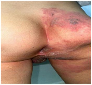
• During our examination, the patient was conscious, eupneic at 19 cpm, normocardial at 89 bpm, apyretic at 37.2°C, with a good staturo-ponderal development, and who presents at the genital level a right unilateral erythematous placard involving 3 metamers: L1-S2-S3 surmounted by multiple vesicles grouped together and other bullae, flaccid with cloudy and hematic content (Figures 1,2). His urine stream was normal, he had no large bursa and presented with sub-centimeter adenopathies at the inguinal level.
• Given this presentation, two diagnoses were evoked: genital shingles and HSV infection, which indicated a consultation with the child psychiatrist and eliminated the possibility of sexual abuse.
• The diagnosis retained was genital shingles in its multimetameric form. In view of this unusual and extensive presentation, a systemic antiviral treatment based on Aciclovir was started (10 mg/kg/8h), associated with a paracetamol analgesic (15 mg/kg/6h), an antihistamine and an antiviral cream associated with an antiseptic solution.


• HIV serology was negative.
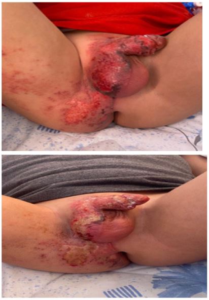
• The evolution was favorable, with healing and the beginning of epidermization (Figure 3).
• The evolution after 2 months of the infectious episode was a total cure with disappearance of the lesions (Figure 4).
Discussion
Humans are the exclusive reservoir of Varicella Zoster Virus (VZV), a neurotropic virus with the peculiarity of remaining quiquiescent in the dorsal sensory ganglia. VZV belongs to the Herpes viridae family, an envelope virus with a DNA genome. It has a particular affinity for the skin, nervous system and lungs [3].
The incidence of shingles increases with age, but children who have had chickenpox in the first year of life (or in utero) have an increased risk of developing shingles [4].
Infection occurs through direct contact with a person with chickenpox (respiratory droplets or vesicle fluid) or a person with shingles (vesicle fluid). Reactivation is favored by immunodepression.
The particularity of our case is its initial exposure in utero and the reactivation in the form of genital shingles.
After primary infection (chickenpox), the virus reaches the sensory ganglia by hematogenous and/or neurogenic route from the skin or mucous membranes. Upon reactivation, it migrates along sensory nerve fibers to the skin. A characteristic vesicular eruption appears in the metamer corresponding to the spinal ganglion colonized during the primary infection. This joins the elementary lesion in our case which shows an inflammatory placard topped by a vesicle following 3 metamers, which is the particularity of our case.
The differential diagnosis can be made with a herpes simplex virus infection which is of sexual transmission. The genital location and the appearance of vesicles that may coalesce into a cluster may raise suspicion of sexual abuse in children. As reported in 2 cases in the literature, in which the diagnosis of certainty was made through the search for antibodies by immunofluorescence to exclude any possibility of sexual abuse. In our case, questioning by the child psychiatrist’s team made it possible to exclude this possibility [5,6].
The first step in the management of the disease is daily bathing, local care with an antiseptic, and oral aciclovir is the first-line treatment at a rate of 10 mg/Kg/8h for 5 to 7 days. Other antivirals such as famciclovir and valacyclovir are not indicated in infants [7,8].
The prognosis was excellent. The most frequent complications were secondary bacterial superinfection, depigmentation and scarring; other complications were rarer, such as encephalitis, ventriculitis, sclerokeratitis and anterior uveitis [9].
Herpes zoster, although rare in healthy children, should also be included in the differential diagnosis of painful genital or anal vesicular rash.
Conclusion
Herpes zoster is a rare condition in children, with a mostly favorable evolution without further consequences, especially pain as reported in the adult form. Its diagnosis is essentially clinical. It does not require specific treatment except for ophthalmic forms, complicated and extensive forms or in case of immunocompromised terrain.
Conflict of interest: None.
References
- Civen R, Chaves SS, Jumaan A, Wu H, Mascola L, Gargiullo P, et al. The incidence and clinical characteristics of herpes zoster among children and adolescents after implementation of varicella vaccination. Pediatr Infect Dis J. 2009; 28: 954‑9.
- Kennedy PGE, Rovnak J, Badani H, Cohrs RJ. A comparison of herpes simplex virus type 1 and varicella-zoster virus latency and reactivation. J Gen Virol. 2015; 96: 1581‑602.
- Fillet AM. Histoire naturelle de l’infection à VZV: Physiopathologie, mécanismes d’action et critères virologiques d’évaluation des antiviraux. Médecine Mal Infect. 1998; 28: 767‑74.
- Feder HMJ, Hoss DM. Herpes Zoster in Otherwise Healthy Children. Pediatr Infect Dis J. 2004; 23: 451.
- Filippova A, Templier I, Lupo J, Leccia MT, Morand P, et al. Genital herpes zoster in an immunocompetent child. Eur J Dermatol EJD. 2020; 30: 54‑5.
- Simon HK, Steele DW. Varicella: Pediatric genital/rectal vesicular lesions of unclear origin. Ann Emerg Med. 1995; 25: 111‑4.
- Levin MJ, Dahl KM, Weinberg A, Giller R, Patel A, et al. Development of Resistance to Acyclovir during Chronic Infection with the Oka Vaccine Strain of Varicella‐Zoster Virus, in an Immunosuppressed Child. J Infect Dis. 2003; 188: 954‑9.
- Acyclovir (Oral Route, Intravenous Route) Proper Use - Mayo Clinic. 2023.
- Kanamori K, Shoji K, Kinoshita N, Ishiguro A, Miyairi I. Complications of herpes zoster in children. Pediatr Int. 2019; 61: 1216‑20.