
Journal of Clinical Images and Medical Case Reports
ISSN 2766-7820
Case Report - Open Access, Volume 4
A case of polycystic liver with cyst infection
Guo Sheng1; Liu Ying1; Liu Changhong1; Liao Yan1; Wei Yingfeng2*
1Department of Severe Hepatopathy, Institute of Liver Disease, Ganzhou Fifth People’s Hospital, Ganzhou City 341000, Jiangxi Province, China.
2Department of Gastroenterology, Ganzhou People’s Hospital, Ganzhou City 341000, Jiangxi Province, China.
*Corresponding Author : Wei Yingfeng
Department of Gastroenterology, Ganzhou People’s Hospital, Ganzhou City 341000, Jiangxi Province, China.
Email: 56476749@qq.com
Received : Aug 29, 2023
Accepted : Sep 18, 2023
Published : Sep 25, 2023
Archived : www.jcimcr.org
Copyright : © Yingfeng W (2023).
Abstract
Objective: A case of polycystic liver infection was reported, and the diagnosis and treatment of polycystic liver infection was summarized based on its clinical features, imaging findings, and the process of diagnosis and treatment, so as to improve doctors’ attention to polycystic liver infection and the level of diagnosis and treatment.
Methods: The clinical data of the patient with polycystic liver infection was retrospectively analyzed.
Results: The patient in this case had fever with abdominal pain as the main clinical manifestation, and was diagnosed with 1. polycystic liver combined with infection 2. polycystic kidney, based on medical history, signs, laboratory and imaging examinations.
Conclusion: Polycystic liver combined with cystic infection is a serious complication, and the diagnosis can be made by combining clinical manifestations, laboratory tests and imaging findings. This complication should be diagnosed and treated early to prevent it from progressing to bacteremia, sepsis or even life-threatening.
Keywords: Polycystic liver disease; Cyst infection.
Citation: Sheng G, Ying L, Changhong L, Yan L, Yingfeng W. A case of polycystic liver with cyst infection. J Clin Images Med Case Rep. 2023; 4(9): 2610.
Clinical information
A 27-year-old woman was admitted to with “fever with abdominal pain, increased abdominal circumference, and loss of appetite for 4 days”. The patient was admitted to the hospital for “fever with abdominal pain, increased abdominal circumference and decreased appetite for 4 days”. 4 days ago, the patient developed fever without obvious triggers, with a maximum temperature of 39.3OC, and was treated with fluids by the local clinic, and the peak of the fever gradually shifted downward, with low-grade fever as the main cause, accompanied by abdominal pain, abdominal circumference increased compared with the previous one, with mid-abdominal area as the main cause, with intermittent severe pain that could be relieved after half an hour, accompanied by decreased appetite, occasional nausea and desire to vomit, without coughing, coughing up sputum, no dizziness, headache and so on. Yesterday, the abdominal pain was aggravated, and the patient was referred to the local Chinese medicine hospital, where the abdominal ultrasound showed polycystic liver and polycystic kidney, and the symptoms did not improve significantly after the treatment of pain relief.
Past history: Denied history of infectious diseases such as tuberculosis, hepatitis and typhoid fever; denied history of hypertension, diabetes mellitus and chronic nephritis; denied history of surgery, trauma and blood transfusion; denied history of allergy to medicines and other foods. Personal history: Born and raised in the same place, farmer, never been to an epidemic area, denies contact with infected water, denies smoking and alcohol consumption. Physical Examination: Body temperature: 36.7OC, pulse: 150 beats/min, respiration: 30 beats/min, blood pressure: 100/60 mmHg No yellow staining of the skin and mucous membranes all over the body, no liver palms or spider nevi. There was no yellowing of the sclera, and the respiratory sounds of both lungs were clear, with no dry or wet rales detected. The heart rhythm was synchronized, the epigastrium was distended, and there were no varicose veins in the abdominal wall. Abdominal muscles were tight, with diffuse pressure and rebound pain throughout the abdomen. A large mass was palpable, hard, ill-defined, with tenderness. The liver was accessible 5 fingers below the ribs (Figure 1), the gallbladder was not accessible, Murphy’s sign was negative, percussion pain in the hepatic region was positive, abdominal mobile turbidities were negative, and there was no edema in both lower limbs.
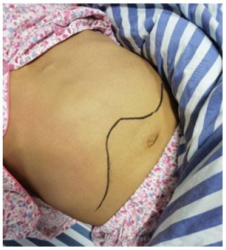


Table 1: Results of liver function and infection indexes during the course of the disease.
| 0* | 3 | 6 | 9 | 16 | |
|---|---|---|---|---|---|
| WBC (*10^9/L) | 14.48 | 18.15 | 20.66 | 13.37 | 10.19 |
| PCT (ng/mL) | 0.844 | 0.598 | 0.323 | 0.115 | 0.206 |
| IL-6 (pg/mL) | 402.32 | 89.53 | 91.23 | 8.94 | 28.54 |
| CRP (mg/L) | 298.6 | 202.5 | 124.8 | 58.0 | 28.3 |
| GGT (U/L) | 391 | 353 | 232 | 176 | 165 |
| ALP (U/L) | 222.2 | 191.9 | 184.2 | 182.4 | 179.6 |
Note: *The day of admission is day 0.
After the patient was admitted to the hospital and the relevant examinations were actively improved, the laboratory results showed that the white blood cell count was 14.48x10^9/L, the percentage of neutrophils was 86.2%, the C-reactive protein was 298.6 mg/L, the calcitoninogen was 0.844 ng/mL, the IL-6 was 402.32 pg/mL, the Total Bilirubin (TBIL), Alanine Aminotransferase (ALT) Total Bilirubin (TBIL), Alanine Aminotransferase (ALT) were normal, Alkaline Phosphatase (ALP) was 222.2 U/L, Gamma-Glutamyltransferase (GGT) was 391 U/L, Alpha-Fetoprotein (AFP), and serum glycoantigen 199 (CA 199) were normal, and blood cultures were negative. Abdominal CT showed diffuse and irregular cystic lesions in the liver (Figure 2). Abdominal CT coronal plane showed an enlarged liver that occupied most of the abdominal cavity (Figure 3). Polycystic Liver Disease (PLD) with hepatic cyst infection was diagnosed and classified as severe phenotype (Gigot type III according to Gigot staging) [1], and was treated with anti-infective therapy of imipenem cilastatin, followed by a step-down to cefoperazone sulbactam sodium, with improvement in the liver function and infection indexes (Table 1), and the infection was controlled, with improvement in the abdominal pain and fever, and the abdominal circumference was reduced compared to the previous one. Considering that the volume of the patient’s normal liver parenchyma was much smaller than the volume of the hepatic cyst, we recommended liver transplantation, but the patient was finally discharged due to his own financial reasons.
Discussion
Polycystic Liver Disease (PLD) is a hereditary disease that often occurs in association with polycystic kidneys, with a prevalence of approximately 1-2% [2]. Most patients with PLD are asymptomatic and do not require treatment. When the enlarged liver compresses the surrounding organs, the corresponding clinical manifestations, such as abdominal distension, abdominal pain, and shortness of breath, can occur, and serious complications such as traumatic rupture, co-infection, and co-haemorrhage can also occur. For patients with severe PLD, liver transplantation is the only treatment.
Cystic infection is a rare complication of polycystic liver, which usually presents as acute pain or pressure and discomfort in the right upper abdomen and right quaternary ribs, and may be accompanied by fever with chills and elevated white blood cells. Its reported mortality rate is about 3% [3,4], so it should be diagnosed and treated early to prevent it from progressing to bacteremia, sepsis or even life-threatening. The current gold standard for the diagnosis of polycystic liver combined with cyst infection is the presence of bacteria and/or neutrophils in the cystic cavity contents [5]. However, due to the limited implementation of cystocentesis, specimen collection is not possible, and in most cases the diagnosis is made by combining clinical manifestations, laboratory tests and imaging findings.
Early diagnostic markers of polycystic liver co-infection include white blood cell count, neutrophil percentage, Erythrocyte Sedimentation Rate (ESR), C Reactive Protein (CRP), and Procalcitonin (PCT), of which CRP and PCT are the two acute serum proteins. CRP and PCT are two acute serum proteins, and the levels of CRP and PCT are associated with the onset, progression, and outcome of sepsis. 51% of patients with PLD have elevated γ-glutamyltranspeptidase, and 17% have elevated serum alkaline phosphatase, both of which may reflect cholangiocyte activation [6]. In patients with polycystic liver, aminotransferases are normal or mildly elevated, and bilirubin is rarely elevated, but may be elevated if the polycystic liver compresses the common bile duct [7].
In recent years, Ultrasonography (US), X-ray Computed Tomography (CT), Magnetic Resonance Imaging (MRI), and Fluorodeoxyglucose Positron Emission Tomography combined with Computed Tomography (18F-FDG PET/CT) imaging have been used to diagnose possible polycystic liver co-infection. Ultrasound is the first choice of examination for polycystic liver, ultrasound can see multiple round or round-like homogeneous non-echoic areas, the cyst wall is clear and curved strong echogenicity; CT polycystic liver shows smooth border, sharp multiple round, round-like watery density foci, uniform density, CT value of 0-20 HU, the average of 14 HU, enhancement of scanning foci of non-enhancement, with the infection of polycystic liver can be accompanied by segregation and small fragments of components, similar to the wall of the capsule enhancement. According to one study, about 40% of patients with liver cysts combined with infection have negative USG and CT results [8]. MRI scanning of polycystic liver has uniform signals, clear borders, low signals on T1WI, markedly high signals on T2WI, and high signals on heavy T2WI. Suwabe T [9] believes that the level of fluid-liquid in polycystic livers and thickening of the cystic wall in MRI are evidence of infection. Diffusion-Weighted Imaging (DWI) is now becoming a valuable MRI technique for the evaluation of renal and hepatic lesions. DWI expresses the rate of diffusion of water molecules between tissues in terms of Apparent Diffusion Coefficients (ADC), and a significant decrease in the ADC value is considered to indicate polycystic liver co-infection. However, the lack of an established ADC threshold for the indication of polycystic liver co-infection limits its use in clinical work [10,11]. 18F-FDG PET/CT is a new imaging technique that helps to identify renal and hepatic cystic infections based on the increased uptake of the radiolabeled glucose analog 18F-FDG by highly metabolically active and inflammatory cells and the absence of hepatic and renal toxicity of 18F-FDG, but due to the high cost and availability, it is not suitable for use in clinical work [11]. Which is not hepato-renal toxic, but is limited by high cost and limited availability.
This case is a middle-aged female patient with fever with abdominal pain and increased abdominal circumference as the main manifestations. Physical examination showed that the liver was 5 cm below the ribs, laboratory tests showed that inflammatory indexes were obviously elevated, GGT and ALP were mildly elevated, and the results of pathogenetic tests were negative, and the CT scan suggested polycystic liver. Although the cystic fluid specimen had not been obtained, the combination of the clinical manifestations, laboratory tests, and the results of the imaging tests could lead to the diagnosis of polycystic liver with cyst infection. It has been reported that the most common pathogen of polycystic liver co-infection is Escherichia coli, followed by Klebsiella spp, Enterobacter spp, and Pseudomonas aeruginosa [2]. However, recent studies have shown that Klebsiella spp. has surpassed Escherichia coli as the most predominant causative agent of polycystic liver co-infections [12]. The infection was controlled and clinical symptoms resolved after treatment with beta-lactams.
In summary, Polycystic liver combined with cystic infection is a serious complication, and the diagnosis can be made by combining clinical manifestations, laboratory tests and imaging findings. This complication should be diagnosed and treated early to prevent it from progressing to bacteremia, sepsis or even life-threatening.
References
- Liu HB. Progress in the structural study of intrahepatic bile duct epithelial cell protocilia. [J] Chinese Journal of Combined Chinese and Western Medicine Surgery. 2009; 2: 199-200.
- Cornec-Le Gall E, Torres VE, Harris PC. Genetic Complexity of Autosomal Dominant Polycystic Kidney and Liver Diseases. J Am Soc Nephrol. 2018; 29: 13-23.
- Reid-Lombardo KM, Khan S, Sclabas G. Hepatic cysts and liver abscess. [J] Surg Clin North Am. 2010; 90: 679-697.
- Lantinga MA, Drenth JP, Gevers TJ. Diagnostic criteria in renal and hepatic cyst infection. [J] Nephrol Dial Transplant. 2015; 30: 744-751.
- Boillot O, Cayot B, Guillaud O, et al. Partial major hepatectomy with cyst fenestration for polycystic liver disease: Indications, short and long-term term outcomes.[J] Clin Res Hepatol Gastroenterol. 2021; 45: 661-670.
- Zhang Aung-chun. Analysis of cases of polycystic liver combined with infection and literature review [D]. Hebei Medical University. 2021.
- Drenth JP, Chrispijn M, Nagorney DM, et al. Medical and surgical treatment options for polycystic liver disease.[J] Hepatology. 2010; 52: 2223- 2230.
- Marten A. Lantinga and others, Diagnostic criteria in renal and hepatic cyst infection, Nephrology Dialysis Transplantation. 2015; 30: 744-751.
- Suwabe T, Araoka H, Ubara Y, et al. Cyst infection in autosomal dominant polycystic kidney disease: causative microorganisms and susceptibility to lipid-soluble antibiotics. [J] Eur J Clin Microbiol Infect Dis. 2015; 34: 1369-1379.
- Suwabe T, Ubara Y, Sumida K, et al. Clinical features of cyst infection and hemorrhage in ADPKD: New diagnostic criteria. [J] Clin Exp Nephrol. 201Jouret F, Lhommel R, Devuyst O, et al.
- Jouret F, Lhommel R, Devuyst O, Annet L, Pirson Y, et al. Diagnosis of cyst infection in patients with autosomal dominant polycystic kidney disease: Attributes and limitations of the current modalities.[J] Nephrol Dial Transplant. 2012; 27: 3746-37512.
- Luo M, Yang X, Tan B, et al. Distribution of common pathogens in patients with pyogenic liver abscess in China: A meta-analysis.[J] European journal of clinical microbiology & infectious diseases: Official publication of the European Society of Clinical Microbiology. 2016; 35: 1557-1565.