
Journal of Clinical Images and Medical Case Reports
ISSN 2766-7820
Case Report - Open Access, Volume 4
Tuberculous spondylitis masked by pyelonephritis
Danay Pérez Mijenes*; Mariana Lobo; Marilia Santos Silva; Joana Alves Vaz
Internal Medicine Department, Hospital Pedro Hispano, Unidade Local de Saúde de Matosinhos, Senhora da Hora, Portugal.
*Corresponding Author : Danay Pérez Mijenes
Internal Medicine Department, Hospital Pedro Hispano, Unidade Local de Saúde de Matosinhos, Senhora da Hora, Portugal.
Email: danaypm@yahoo.es
Received : Sep 02, 2023
Accepted : Sep 19, 2023
Published : Sep 26, 2023
Archived : www.jcimcr.org
Copyright : © Mijenes DP (2023).
Abstract
Tuberculosis can affect any organ and the spine is the most affected bone structure in the case of musculoskeletal tuberculosis (TB). In this article, we present a case of an 87-year-old woman with chronic low back pain and previous contact with pulmonary tuberculosis. She was admitted to hospital for decompensated heart failure due to uncomplicated acute pyelonephritis without microbiological agent identification. She maintained persistent fever, low back pain and elevated inflammatory parameters despite antibiotics, so other etiologies have been thought. Magnetic resonance showed tuberculous spondylitis and she started anti-tuberculous (anti-TB) therapy that must be complied with for 12 months. This case highlights the importance of a high index of clinical suspicion as a requirement for a correct and prompt diagnosis.
Keywords: Spinal tuberculosis; Psoas abscess; Secondary immune thrombocytopenia.
Citation: Mijenes DP, Lobo M, Silva MS, Vaz JA. Tuberculous spondylitis masked by pyelonephritis. J Clin Images Med Case Rep. 2023; 4(9): 2612.
Introduction
Tuberculosis (TB) is caused by Mycobacterium tuberculosis and is one of the oldest diseases in the world [2]. Spinal TB is the most common musculoskeletal manifestation, affecting about 1 to 2% of all cases of TB. Percival Pott described the associated paraplegia in 1779 due to destruction of the anterior spinal column and progressive kyphosis. The onset of spinal TB is insidious, typically progressing over four to 11 months [1] and is the result of haematogenous dissemination from a primary focus [2]. A biopsy of the pathology is mandatory to acquire tissue or pus to differentiate the lesion from tumour and other infections. Culture of Mycobacterium tuberculosis remains the benchmark for definitive diagnosis and allows determination of sensitivity to antibiotics [2].
Case description
We present an 87-year-old woman with medical history of arterial hypertension, dyslipidemia and osteoarthritis associated with chronic low back pain. She revealed a weight loss of 5 kg in the last 6 months. She mentioned contact with her husband who had had pulmonary tuberculosis for more than 30 years. At that time, she performed screening tests that were negative. Admitted to the emergency department with fever, urinary symptoms (dysuria and pollakiuria) and dyspnea with 5 days of evolution. At observation was hypertensive (systolic blood pressure 180 mmHg), polypneic and tachycardic.
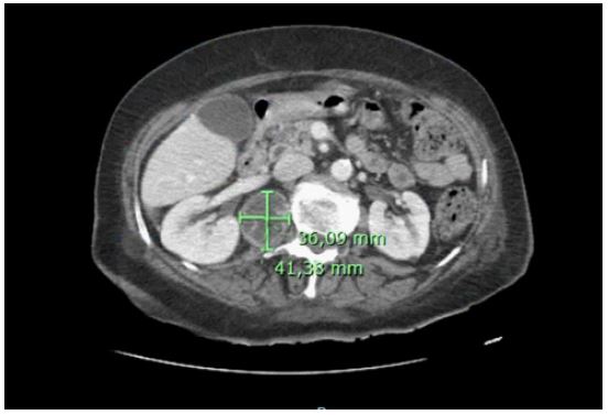
Blood tests revealed elevated inflammatory parameters (C-reactive protein (CRP) 131 mg/L, leukocytes 13630/μl and erythrocyte sedimentation rate (ESR) 66 mm/h), NT-proBNP 3200 pg/mL and urinary sediment with leukocyturia (97/field). Decompensated acute heart failure in the setting of uncomplicated acute pyelonephritis was assumed and empirical antibiotic therapy with ceftriaxone was started. The urine culture was non-valuable. She developed persistent fever and high inflammatory parameters with no other clinical signs of infection except dysuria and discontinued ceftriaxone after 4 days and started piperacillin/tazobactam. All blood cultures, urine cultures and viral serologies, including HIV, were negative. Urine cytology was negative for malignant cells and also for mycobacterium tuberculosis PCR. The investigation was extended by a thoraco-abdominopelvic CT scan which demonstrated an abscess of the right iliopsoas muscle (41 mm x 36 mm) (Figure 1), with destruction of the L1 vertebral body. She was submitted to CT-guided percutaneous drainage of the abscess. Microscopy revealed acid-fast bacilli, positive nucleic acid amplification test for M. Tuberculosis and growth of M. Tuberculosis multisusceptible in culture exam. Magnetic resonance imaging (MRI) of the spine confirmed the infectious process of D12-L1 (Figure 2), with endocanal involvement, without compressive myelopathy. There were no neurological deficits or signs of spinal cord involvement throughout hospitalisation. Active pulmonary involvement was excluded with thoracic CT without changes and negative sputum mycobacteriology (more than 3 samples). Screening of serial urine samples was negative for Mycobacterium tuberculosis.
Tuberculous spondylitis of D12-L1 was assumed and treatment with rifampicin, isoniazid, pyrazinamide and ethambutol was started, without surgical indication.
After 12 days of initiation of TB drugs, she developed severe secondary immune thrombocytopenia, with platelet counts less than 1x10³/μl, with weakly positive platelet antibodies.
Although thrombocytopenia was more related to to rifampicin and all anti-TB were stopped. She was treated with 2 days of intravenous immunoglobulins with improvement in platelet count. Reinitiation of ethambutol, given the lower risk of thrombocytopenia, was associated with a new drop in platelet counts and a new cycle of immunoglobulins, assuming failure of the first cycle. Pyrazinamide and isoniazid was sequentially introduced after platelet count stabilization, and finally, instead of rifampicin, levofloxacin was started. There was clinical and analytical improvement and started lift with Jewett vest due to complaints of low back pain. She was discharged to home, under oral antibacillary drugs for follow up in infectious diseases consultation with imaging reassessment after 3 months. Indication to complete with anti-tuberculosis therapy for 12 months.

Discussion
Tuberculosis can affect any organ, with musculoskeletal involvement occurring in 10% of cases of extra-pulmonary tuberculosis [1]. More than 50% of cases occur in the spine, most frequently in lumbar or thoracic spine [1]. Pott’s disease results from reactivation of a bacillus in bone, which will have become lodged during a mycobacteremia in the primary infection [2].
The clinic is non-specific and insidious and can often go unnoticed [1,2]. Chronic low back pain is a common symptom and one third of patients have constitutional symptoms or fever [2]. So it is necessary to review epidemiological previous contacts with tuberculosis [1], as blood tests and spinal X-rays may be normal or unremarkable in their results in the early stages, with X-ray changes only appearing after 30% loss of bone mass [1,2]. The use of CT allows visualization of lesions at an earlier stage and is a good tool for percutaneous guided biopsy [1], although MR imaging is the recommended imaging method for assessing soft tissue, spinal cord and bone involvement [1]. Complications arise from the delay in diagnosing spondylitis. This explains why neurological involvement is quite common, affecting 23 to 76% of patients [1,2]. Paraplegia, one of the most feared neurological disorders, occurs in 10 to 30% of patients [3]. The abscesses results from a contiguity solution with the spondylitis, normally have a paravertebral location and are described as “cold”, without signs of inflammation [1,3].
The diagnosis takes place by microbiological identification [1]. The acid fast bacilli can be positive in 25 to 75% of cases [3]. Ziehl-Nielsen staining can detect mycobacteria but not distinguish between species [1]. Cultural growth is the gold standard for the diagnosis of tuberculosis but can take several weeks to positive [1]. Molecular methods, for example, polymerase chain reaction test amplifies the M. Tuberculosis DNA in the sample [1]. The Xpert MTB in 2 hours can identifies M. Tuberculosis and even resistance to rifampicin [1].
Treatment includes anti-TB therapy and, depending on each case, surgical treatment [1]. The first-line regimen is rifampicin, ethambutol, isoniazid, and spretomycin for 2 months, followed by isoniazid, rifampicin, and ethambutol for 10 months [3]. The duration of the treatment should be determined according to the patient’s evolution and the imaging reassessment [1]. There are no guidelines for surgical indications but progressive neurological deficits, progressive spinal deformity, medical treatment failure and diagnostic uncertainty seem to be the most frequent indications [1,5].
This case presents an elderly patient with degenerative osteoarticular pathology who was hospitalized for decompensated heart failure and whose clinical and analytical findings were suggestive of pyelonephritis, which developed unfavourably and culminated in a diagnosis of tuberculous spondylitis. In this case, a correct history focused on the epidemiological context and imaging tests were essential for the detection of a lumbar infectious process and a local complication, as well as for the collection of microbiological products. The positivity of the molecular tests allowed early initiation of antituberculostatic drugs and reduced the likelihood of later complications. As there were no focal neurological signs or other complications, treatment consisted only of anti-TB. However, pharmacological treatment ended with one complication, thrombocytopenia, which was associated with the use of rifampicin. It is important to remember that this treatment is not harmless and that one should be even more attentive in elderly and polymedicated patients.
Conclusion
In a patient with a suggestive epidemiological and clinical context, a high level of suspicion is required to make a rapid diagnosis. If this infection is not promptly diagnosed and treated, multiple complications, including paraplegia, bacteremia and death, may result. Imaging tests such as MRI and CT help diagnose the disease, but a microbiological diagnosis is needed for confirmation. The earlier the diagnosis is made, the greater the chance of a successful outcome.
References
- Leowattana W, Leowattana P, Leowattana T. Tuberculosis of the spine. World J Orthop [Internet]. 2023; 14(5): 275-93. Available from: http://dx.doi.org/10.5312/wjo.v14.i5.275
- Pigrau-Serrallach C, Rodríguez-Pardo D. Bone and joint tuberculosis. Eur Spine J [Internet]. 2013; 22(S4): 556-66. Available from: http://dx.doi.org/10.1007/s00586-012-2331-y
- Jain AK, Rajasekaran S, Jaggi KR, Myneedu VP. Tuberculosis of the spine. J Bone Joint Surg Am [Internet]. 2020; 102(7): 617-28. Available from: http://dx.doi.org/10.2106/jbjs.19.00001
- Coughlan CH, Priest J, Rafique A, et al. Spinal tuberculosis and tuberculous psoas abscess. BMJ Case Rep 2019; 12: e233619. doi:10.1136/bcr-2019-233619
- Ferrer MF, Torres LG, Ramírez OA, Zarzuelo MR, del Prado González N. Tuberculosis of the spine. A systematic review of case series. Int Orthop [Internet]. 2012; 36(2): 221-31. Available from: http://dx.doi.org/10.1007/s00264-011-1414-4