
Journal of Clinical Images and Medical Case Reports
ISSN 2766-7820
Clinical Image - Open Access, Volume 4
An uncommon cold: A boy with a runny nose
*Corresponding Author : Chee Teck Koh
Department of Paediatrics, Khoo Teck Puat- National University Children’s Medical Institute, National University Health System, Singapore.
Email: kct0831@gmail.com
Received : Sep 01, 2023
Accepted : Sep 20, 2023
Published : Sep 27, 2023
Archived : www.jcimcr.org
Copyright : © Chee Teck K (2023).
Citation: Chee Teck K. An uncommon cold: A boy with a runny nose. J Clin Images Med Case Rep. 2023; 4(9): 2616.
Case presentation
A 4-year-old boy with autism spectrum disorder presented to the emergency department complaining of 4 days of fever, runny nose and a mild cough. Examination was unremarkable. He was diagnosed with a viral upper respiratory tract infection and discharged. He returned 48 hours later with persistent fever, right purulent nasal discharge and unilateral facial swelling.
Clinically, there was mucopurulent nasal discharge from the right nostril with unilateral infraorbital facial swelling. There was marked tenderness extending from the right infraorbital region to the right nasal bridge. The ophthalmological examinations were unremarkable.
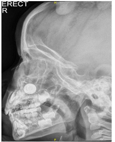
There was a concern of rhinosinusitis with possible right maxillary osteomyelitis, and a radiograph of the paranasal sinuses was done. The antero-posterior radiograph of paranasal sinuses (Figure 1) showed a round radio-opaque foreign body in the right nostril. The lateral radiograph of paranasal sinuses (Figure 2) showed a typical double ring or halo sign that was suggestive of a button battery in the right nostril, without radiological finding of facial osteomyelitis. The diagnosis was right rhinosinusitis with right nasal button battery.
He underwent removal of right nasal button battery and debridement urgently. Intra-operative examination revealed necrotic tissue involving the right anterior inferior turbinate, anterior half of the septum and nasal floor. There was no septal perforation. Post operatively, he was treated with oral co-amoxiclav for 1 week and underwent repeated examination in the first month to assess the recovery. He recovered well and was subsequently discharged after 2 months.
Button battery impaction in the nasal cavity is an emergency and leads to serious complications such as septal perforation, necrosis of tissue mucosa and facial cellulitis. The possible mechanisms of these are liquefactive necrosis of corrosive battery content, production of local current, direct current burn and pressure necrosis of impacted foreign body [1,2]. It has a characteristic appearance on radiography: they have a bilaminar structure; so they appear as a double ring or halo (double density) on anteroposterior view and a step-off at the separation between the anode and cathode on lateral view [2,3]. Our patient’s plain radiograph demonstrates the characteristic appearance of nasal button battery.

This case illustrates the importance of screening for foreign body in the case of unilateral purulent nasal discharge especially in cases with limited history.
Declaration of competing interest: The authors have no conflicts of interest relevant to this article.
References
- Loh WS, Leong JL, Tan HK. Hazardous foreign bodies: complications and management of button batteries in nose. Ann Otol Rhinol Laryngol. 2003; 112: 379-383.
- Bhatia R, Singhal SK, Dass A. Button Cell Causing Septal Perforation in a Child. Clinical Rhinology: An International Journal, 2010; 3(3): 161-163.
- Kaur J, Ravikumar R, Viswanatha B, Vijayashree MS. An Interesting Case of Button Battery Causing Septal Perforation. Research in Otolaryngology. 2014; 3(6): 89-91.