
Journal of Clinical Images and Medical Case Reports
ISSN 2766-7820
Research Article - Open Access, Volume 4
miR-30a-3p/IGF2BP1 affect the proliferation and migration of lung adenocarcinoma cells
Jing Xu†; Feng Wang†; Machicheng Bao†; Jia Liu*
Institute of Neuroscience and Animal Zoology Department, Kunming Medical University, Kunming, 650031, China.
†These Authors Contributed Equally.
*Corresponding Author : Jia Liu
Institute of Neuroscience and Animal Zoology Department, Kunming Medical University, Kunming, 650031, China.
Email: liujiaaixuexi@126.com
Received : Sep 08, 2023
Accepted : Sep 27, 2023
Published : Oct 04, 2023
Archived : www.jcimcr.org
Copyright : © Liu J (2023).
Abstract
Background: IGF2BP1 is a carcinomatous fetal protein associated with proliferation, migration and invasion of tumor cells. The different roles of miR-30a-3p as a tumor suppressor or an oncogene depend on the target genes it regulates.
Methods: Downloading the relevant data of lung adenocarcinoma from the TCGA database for bioinformatics analysis to determine the key gene IGF2BP1 and its miRNA that affect the progression of lung adenocarcinoma, and using qPCR to verify their expression and correlation in lung adenocarcinoma. Gene enrichment analysis of IGF2BP1 related pathways, and the function of miR-30a-3p/IGF2BP1 axis in lung adenoma was verified by CCK-8 detection, MTT assay, double luciferase gene reporting assay, cell cloning assay and wound healing assay.
Results: As predicted, IGF2BP1 was highly expressed in lung adenocarcinoma tissues, affecting TNM stage and survival status. In addition, IGF2BP1 was negatively correlated with the expression of miR-30a-3p, and the low expression of miR-30a-3p had a poor survival period. Luciferase result confirmed the existence of a targeting relationship between miR-30a-3p and IGF2BP1. Gene enrichment analysis showed that the high expression of IGF2BP1 was correlated with the proliferation of lung adenocarcinoma cells. The experimental results of this study showed that cell viability, proliferation and migration of cells were decreased in the si-IGF2BP1 group and miR-30a-3p-mimics group. In the wound healing assay, compared with the mimics group, the migration ability of cells in the mimics+oe-IGF2BP1 group was improved, which was consistent with the results in the control group.
Conclusion: miR-30a-3p can affect the activity of lung adenocarcinoma cells and promote cell proliferation via regulating IGF2BP1. The miR-30a-3p/IGF2BP1 axis plays an important role in the progression of LUAD and can be used as a potential therapeutic target.
Keywords: Lung adenocarcinoma; IGF2BP1; Proliferation; Migration; miR-30a-3p.
Citation: Xu J, Wang F, Bao M, Liu J. miR-30a-3p/IGF2BP1 affect the proliferation and migration of lung adenocarcinoma cells. J Clin Images Med Case Rep. 2023; 4(10): 2628.
Introduction
At present, lung cancer is one of the most dangerous malignant tumors to human health and life [1], accounting for an important proportion of cancer deaths worldwide, and lung adenocarcinoma is the most common type of lung cancer [2]. Compared with other lung cancer subtypes, the prevalence of lung adenocarcinoma has gradually increased, and has occupied the first place of all lung cancer pathological types [3]. The methods of prevention and treatment of lung cancer at home and abroad also emerge endlessly. However, its early diagnosis, treatment and prognosis have not been significantly improved.
IGF2BPs are insulin-like growth factor-2 mRNA binding proteins, including IGF2BP1, IGF2BP2 and IGF2BP3, which are a highly stable protein family [4]. IGF2BPs is also a post-transcriptional fine-tuning factor involved in the regulation of tumor cell proliferation, survival, and metastasis. IGF2BP1 and IGF2BP3 are carcinofetal proteins, which are associated with poor prognosis and metastasis of various human cancers [5]. Recent studies have found that IGF2BP1 can play an important role in normal cell proliferation and growth as well as tumor cell apoptosis, migration and invasion by regulating newly discovered cancer-related mrnas (PTEN, ACTB, MAPK4, MKI67, c-MYC and CD44) [6], such as: liver cancer, endometrial cancer, breast cancer, lung cancer, ovarian cancer, squamous skin cancer, thyroid cancer and glioma [7-14]. Therefore, IGF2BP1-mediated cell signaling will become a potential target for cancer therapy [6].
miRNA are a class of small endogenous non-coding RNA that negatively regulate post-transcriptional gene expression by directly degrading mRNA or inhibiting its translation [15]. Although its potential biological function is not completely clear, it can play an important role in cell proliferation, growth, apoptosis and differentiation by regulating the expression of target genes [16-18]. miRNA can promote the invasion and migration of cancer stem cells and regulate the tumor microenvironment. As a member of the human miRNA family, miR-30a has tumor inhibition effects in different types of malignant tumors, such as gastric cancer, renal cell carcinoma, hepatocellular carcinoma, colorectal cancer and bladder cancer [19-23]. miR-30a-3p is a member of the miR-30 family, and its different role as a tumor suppressor or oncogene depends on the target gene in the specific type of cancer. Therefore, it is necessary to clarify the selective role of miR-30a-3p in cancer regulation in order to find more effective treatment methods for cancer. Therefore, it is very important to further explore the role of miR-30a-3p in the pathogenesis of lung cancer.
With the rapid development of genetics, molecular biology and bioinformatics, the target genes and potential regulated mirnas of lung adenocarcinoma can be screened based on the gene expression levels in tumor samples, which plays an important role in further understanding the development process and prognosis of lung adenocarcinoma. In this study, we identified the expression characteristics of IGF2BP1 and its effect in LUAD through bioinformatics analysis, and explored the potential mechanism of miR-30a-3p involved in the regulation of IGF2BP1 and its basic biological function in lung adenocarcinoma.
Methods
Download and analysis TCGA database data
Downloading the TCGA-LUAD data from the TCGA database, and using the 4.1.1 R language software package to process the downloaded data, and obtaining the differentially expressed genes in lung adenocarcinoma. According to the survival curve, IGF2BP1 was screened, and using GEPIA [24] to search the expression level of target gene IGF2BP1 in different parts of normal human body and tumor tissues. The relationship between IGF2BP1 expression and TP53 mutation was studied by cBioPortal for Cancer Genomics [25].
Prediction of miRNA regulating IGF2BP1
As the same method, we used lung cancer miRNA-Seq data and download from the TCGA database to analyze their differential expression and screen out down-regulated miRNA. Through whether its survival curve is meaningful, miR-30a-3p regulating IGF2BP1 in lung adenocarcinoma can be finally screened out.
Gene enrichment analysis
We used TCGA-LUAD data for GSEA analysis to explore pathways related to IGF2BP1 expression levels in lung adenocarcinoma. Download the required database files from KEGG database (c2. Cp. KEGG. V7.1. Symbols. GMT) and annotation file, and calculate the NES (enrichment of standardized scores), analysis of gene sets p values. The smaller the p value is, the better the enrichment accuracy is. The greater the absolute value of NES, the smaller the FDR value, and the higher the reliability of the analysis results. If p value and q value of FDR were both lower than 0.05, the gene was significantly enriched.
Quantitative polymerase chain reaction (qPCR)
With the approval of the Pathology Department of the First Affiliated Hospital of Kunming Medical University, paraffin blocks of lung adenocarcinoma tissue and paraffin blocks of normal lung tissue were collected from 14 cases respectively for PCR verification. The wax block tissues of each group were dewaxed and Trizol was added to crack tissues. Total RNA was extracted from tissues, then cDNA was synthesized by reverse transcription and amplified by qPCR. Using 2-ΔΔCt to calculate the expression level of IGF2BP1 gene in lung adenocarcinoma.
A549 cell culture
Selecting human lung cancer cell line A549. The cell line was constructed by D.J. Gerad through lung cancer tissue transplantation culture. A549 cells were cultured with DMED medium containing 10% fetal bovine serum at 37OC and 5% CO2. When the growth of A549 cells reached 80%-90%, cells in good growth condition during logarithmic growth stage were selected and re-inoculated in the culture dish in a ratio of 1 to 3 for further experimental operation.
Experimental grouping
CCK-8 assay and MTT assay were divided into normal group, miR-30a-3p-mimics-NC group, miR-30a-3p-mimics group, miR-30a-3p-instigator-NC group, miR-30a-3p-inhibitor group, si-IGF2BP1-NC group and si-IGF2BP1 group, each group had 6 multiple pores. Colony formation assay was divided into normal group, si-IGF2BP1-NC group and si-IGF2BP1 group. Wound healing assay was divided into normal group, si-IGF2BP1-NC group, si-IGF2BP1 group, control group, miR-30a-3p-mimics group and mimics+oe-IGF2BP1 group, with 3 multiple wells in each group. The luciferase experiment was divided into IGF2BP1-WT-mimics-NC group, IGF2BP1-WT-miR-30a-3p-mimics group, IGF2BP1-MUT-mimics-NC group and IGF2BP1-MUT-miR-30a-3p-mimics group, each group had 6 multiple pores.
CCK8 assay
Logarithmically grown A549 cells were digested with pancreatic enzymes, re-suspended and counted using complete culture medium. Each 96-well plate was 100 μl complete medium with 6 Wells per group. The 96-well plates were cultured in a cell incubator for 24 h. 10 μL CCK-8 reagent was added 4 h before termination of culture, and 96-well plates were put into enzyme marker 4 h later to detect OD value.
MTT assay
96-well plates with a certain cell density were placed into a cell incubator for 24 h culture. Add 20 ul of MTT solution to each well. Incubation continued for 4 h and culture was terminated. Add 150 ul DMSO to each well and shake for 10 min to fully melt the crystals. The wavelength of 490 nm was selected, the light absorption value of each hole was measured on the enzyme marker, and the results were recorded and counted.
Colony formation assay
The density of 1000 cells per well was inoculated into 6-well plates, and the cells were transfected after adherent. After 15 days, cell clone balls appeared. The cells were fixed with 4% paraformaldehyde and stained with crystal violet solution. Finally, the residual liquid was washed with PBS and dried upside down. The number of cell clones was photographed and counted.
Wound healing assay
The cells were inoculated in 6-well plates and cultured for 24 h. Drawing the lines with the pipette tip of a 10 μl pipette perpendicular to the 6-well plate. After lines were drawn, cleaning the cell fragments with PBS and transfected into groups. After transfection, the culture was continued for 48 h and microscopically photographed at 0h, 24 h and 48 h, respectively.
Double luciferase gene reporting experiment
There were two transfection groups, namely IGF2BP1-MUT group (mutant deletion of miR-30c-2-3p binding site) and IGF2BP1-WT group (wild type), the transfection volume was 0.1 μg in each group. 6 multiple wells were set up in each group. After transfection of A549 cells in the above groups for 24 h, the luciferase activity was detected according to the instructions of luciferase kit, and the statistical data were analyzed.
Statistical analysis
Using T test and Chi-square test to evaluate the expression of IGF2BP1 in different clinical samples and the correlation between IGF2BP1 expression and clinicopathological parameters. Receiver operating characteristic (ROC) was used to determine the diagnostic value of IGF2BP1 in lung adenocarcinoma. Using log-rank test to analyze the survival curves of high and low expression IGF2BP1. Single-factor analysis of variance was used for comparison of multiple groups, and independent sample T test was used for data analysis of two groups. All statistical analyses were performed using SPSS 13.0 software (SPSS, Inc., Chicago, IL, USA), and p< 0.05 was considered statistically significant.
Results
High expression of IGF2BP1 in lung adenocarcinoma affects tumor progression
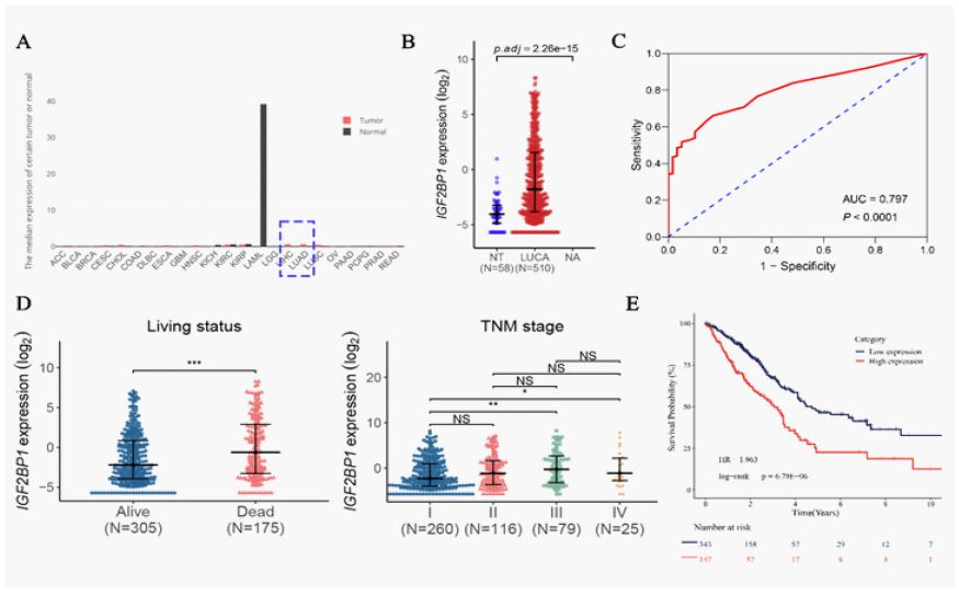
The mRNA expression levels of IGF2BP1 in some normal human tissues and lung adenocarcinoma tissues were verified by GEPIA (Figure 1A). After removing duplicates and unqualified samples, obtaining 58 normal tissue samples and 510 tumor tissue samples. Results showed that IGF2BP1 was low expression in normal tissues and overexpressed in lung adenocarcinoma tissues (Figure 1B). ROC curve suggested that AUC=0.797, P < 0.0001, confirming the importance of IGF2BP1 high expression in the diagnosis of lung adenocarcinoma (Figure 1C). In order to investigate the clinical significance of IGF2BP1 expression in lung adenocarcinoma, 510 patients with lung adenocarcinoma were divided into two groups with high expression of IGF2BP1 and low expression of IGF2BP1. As shown in Table 1 below, there were statistically significant differences in TNM stage, survival status and disease status between the two groups (P< 0.05). In terms of survival, patients with lung adenocarcinoma with high IGF2BP1 expression had a higher mortality rate (P< 0.001) and affected tumor TMN staging (Figure 1D), and lung adenocarcinoma patients with high IGF2BP1 expression had lower survival rates (Figure 1E).
Prediction of miRNA regulating IGF2BP1 in lung adenocarcinoma
The survival of lung adenocarcinoma at 1,3, and 5 years after surgery was shown in Figure 2A, and the predicted results were consistent with the actual observation. We used linear regression analysis and R language to analyze TCGA-LUAD data to explore the potential mechanism of IGF2BP1 affecting lung adenocarcinoma. IGF2BP1 amplification was performed in 9 LUAD patients (n=512), and 222 had IGF2BP1 copy number gain. And the high expression of IGF2BP1 in lung adenocarcinoma was significantly correlated with IGF2BP1 copy number gain (Figure 2B). More importantly, linear regression analysis showed that IGF2BP1 expression was significantly correlated with its methylation level in lung adenocarcinoma (Figure 2C). Using the same method, it is concluded that miR-30a-3p can regulate IGF2BP1 and play a role in lung adenocarcinoma. miR-30a-3p was lower expression in lung adenocarcinoma tissues (n=510) compared with normal lung tissues (n=45) (Figure 2D). In addition, IGF2BP1 expression was negatively correlated with miR-30a-3p expression in lung adenocarcinoma (Figure 2E), and lung adenocarcinoma patients with low miR-30a-3p expression had poorer survival (Figure 2F).
IGF2BP2 affects the proliferation of lung adenocarcinoma cells
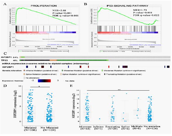
Using the GSEA database to explore the potential biological function of IGF2BP1 up-regulation in lung adenocarcinoma, and finding the target gene significantly enrich in the “proliferation” and “p53 signaling” pathways (Figure 3A,B). As shown in Figure 3C, heat maps of IGF2BP1 expression were searched in cBioPortal for Cancer Genomics, and it was found that high expression of IGF2BP1 in lung adenocarcinoma was correlated with TP53 mutation. The TP53 tumor suppressor gene is frequently mutated in human cancers, and its mutation is often associated with poor prognosis in some cancers. By evaluating the association between IGF2BP1 expression and TP53 mutation in lung adenocarcinoma, we found that IGF2BP1 expression was significantly higher in the TP53 mutant group (Figure 3D). As shown in Figure 3E, the expression of IGF2BP1 in TP53 mutation and TP53 wild type group was compared, and the difference was statistically significant.


IGF2BP1 affects the activity, proliferation and migration of lung adenocarcinoma cells
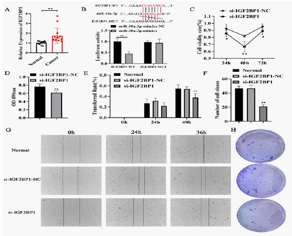
order to verify whether IGF2BP1 is highly expressed in lung adenocarcinoma, using qPCR assay to verify the expression level of IGF2BP1 in normal lung tissues and lung adenocarcinoma tissues. It was found that IGF2BP1 was highly expressed in lung adenocarcinoma tissues compared with normal lung tissues (Figure 4A), which was consistent with the predicted results. According to bioinformatics analysis, miR-30a-3p can play a role in lung adenocarcinoma by regulating IGF2BP1, which was verified. According to the targeting binding site of IGF2BP1 and miRNA miR-30a-3p, double luciferase gene reporting experiment was conducted (Figure 4B), and it was found that there was indeed a targeting binding relationship between miR-30a-3p and IGF2BP1. Gene enrichment analysis showed that IGF2BP1 was associated with the proliferation of lung adenocarcinoma cells, so the interference fragment of IGF2BP1 was constructed. As shown in Figure 4C, the results of CCK8 and MTT experiments showed that the cell vitality and proliferation ability of si-IGF2BP1 group were significantly decreased at 48h (Figure 4D). The results of Wound healing assay (Figure 4E) and cloning experiment (Figure 4F) showed that the proliferation and migration ability of si-IGF2BP1 group were significantly reduced (Figure 4G,H). These results confirm that IGF2BP1 is highly expressed in lung adenocarcinoma, which affects the activity of lung adenocarcinoma cells and promotes the proliferation and migration of A549 cells.
miR-30a-3p affects the proliferation and migration of lung adenocarcinoma cells via regulating IGF2BP1
As shown in Figure 5A, compared with the control group, the cell viability and proliferation capacity of the miR-30a-3p mimics group was decreased (Figure 5B). The wound healing assay results showed that the cell migration ability of the miR-30a-3p mimics group was relatively poor, but the cell migration ability was consistent with that of the control group after transfection of miR-30a-3p mimics with oe-IGF2BP1 treatment (Figure 5C,D). These results indicate that miR-30a-3p can indeed affect the activity, proliferation and migration of lung adenocarcinoma cells by regulating IGF2BP1, thus interfering with the progression of LUAD.
Discussion
The occurrence and development of tumor is the process of abnormal cell proliferation, differentiation, invasion and migration. Despite advances in the treatment of lung cancer in both surgery and medicine, and the precision of lung cancer screening, lung cancer remains one of the cancers with a very high mortality rate. Lung adenocarcinoma is the most common histological subtype of primary lung cancer, and its incidence has exceeded that of other pathological types of lung cancer [1]. Therefore, it is increasingly important to deeply understand the pathophysiology, disease course, diagnosis and prognosis of lung adenocarcinoma.
IGF2BP1 can enhance the viability of tumor cells, and thus play a role in promoting tumor cell growth, migration and invasion. IGF2BP1, a member of IGF2BPs, controls mRNA transport and translation during the development of normal and cancer cells [26]. Gene enrichment analysis showed that IGF2BP1 was associated with signaling pathways such as “proliferation” and “p53 signaling” [27]. Tumor suppressor gene TP53 is the most commonly mutated gene in lung cancer [26]. By evaluating the association between IGF2BP1 expression and TP53 mutation in lung adenocarcinoma, it was found that IGF2BP1 overexpression was correlated with TP53 mutation. Considering the genomic mutation results and the enrichment of the “p53 signaling pathway” in GSEA, it further suggests that there may be a potential association between IGF2BP1 overexpression and TP53 mutations in lung adenocarcinoma. GSEA results showed that TP53 mutations are associated with cell cycle, DNA replication and DNA repair, and TP53 mutations can accelerate cell cycle and DNA replication, thus increasing mutation rate. Therefore, it can be considered that the mutation of TP53 induces DNA replication and DNA damage repair, and the change of related gene IGF2BP1 leads to the occurrence of lung adenocarcinoma. The results of this study showed that the cell vitality, proliferation and migration of cells were significantly decreased in si-IGF2BP1 group, and the number of cell proliferation was decreased. These results indicate that high expression of IGF2BP1 can affect the activity of lung adenocarcinoma cells and promote the proliferation and migration of A549 cells. It has been reported that IGF2BP1 promotes apoptosis of lung cancer cells and thus inhibits proliferation by regulating Netrin-1 [28]. Fang XY [29] et al. also confirmed that IGF2BP1/UHRF2 axis mediated by miR-98-5p promoted the proliferation and inhibited the apoptosis of esophageal squamous cell carcinoma. This is consistent with our experimental results.
It has been reported that the let-7 miRNA family inhibits tumorigenesis by interfering with the synthesis of oncogenic factors, while IGF2BP1 promotes tumorigenesis by interfering with miRNA encoding mRNA in cancer cells [30-32]. mir-30a-3p belongs to the miR-30a family and is involved in affecting the process of many tumors, such as esophageal cancer, cervical cancer, bladder cancer, breast cancer, etc [33-36]. In order to further study the changes of miR-30a-3p expression in lung adenocarcinoma tissues, we analyzed the expression of IGF2BP1 and the miRNA regulating IGF2BP1 in lung adenocarcinoma tissues using R language and the profile data of lung adenocarcinoma expression in the TCGA database. It was screened that miR-30a-3p was involved in regulating the expression of IGF2BP1 in lung adenocarcinoma. Finally, it was found that the expression of miR-30a-3p was significantly reduced and the expression of IGF2BP1 was significantly increased in lung adenocarcinoma tissue. The results of this study suggest that IGF2BP1 overexpression affects stage grading and survival of lung adenocarcinoma. We found that low expression of miR-30a-3p was also associated with poor survival of LUAD, and there was a negative correlation between miR-30a-3p and IGF2BP1 in lung adenocarcinoma. In order to verify this conclusion, according to the targeting binding sites of IGF2BP1 and miRNA miR-30a-3p, double luciferase gene reporting experiments of IGF2BP1 and miR-30a-3p were conducted, and it was found that there was indeed a targeting binding relationship between miR-30a-3p and IGF2BP1. The experimental results showed that the cell viability of miR-30a-3p mimics group was decreased, and the cell proliferation ability was also decreased. Compared with the control group, the cell migration ability of the miR-30a-3p mimmimics group was relatively poor, but the cell migration ability was consistent with that of the control group after transfection with miR-30a-3p mimics and oe-IGF2BP1 treatment. Therefore, we believe that miR-30a-3p can affect the progression of lung adenocarcinoma via regulating IGF2BP1.
Conclusion
In conclusion, through a series of bioinformatics analysis of lung adenocarcinoma, we explored and discussed the important role of miR-30a-3p/IGF2BP1 axis in the progression and prognosis of LUAD. IGF2BP1 gene is enriched in the “proliferation” and “p53 signaling” pathways. miR-30a-3p is associated with IGF2BP1 in lung adenocarcinoma. The above experimental results confirm that miR-30a-3p can indeed affect the activity, proliferation and migration of lung adenocarcinoma cells through the regulation of IGF2BP1, and inhibiting the development of tumors, improving the poor prognosis of lung adenocarcinoma, providing new targets for tumor treatment and new ideas for the study of tumor pathogenesis.
Declarations
Author contribution: Jia Liu: review and editing (equal). Jing Xu: Conceptualization (lead); writing – original draft (lead); formal analysis (lead); writing – review and editing (equal). Feng Wang: Methodology (lead); Software (lead). Machicheng Bao: Conceptualization (supporting); data curation (equal); methodology (equal). All authors have read and agreed to the published version of the manuscript.
Acknowledgement: The study was not funded in any way.
Statements and declarations: The authors have no competing interests to declare that are relevant to the content of this article.
References
- Wang, J, Z. He, and R. Duan. Expression of ASPM in Lung Adenocarcinoma and Its Relationship with Development and Prognosis. Zhongguo Fei Ai Za Zhi. 2020; 23(1): 29-35.
- Balzer, B.W.R, et al. Adenocarcinoma of the Lung in Childhood and Adolescence: A Systematic Review. J Thorac Oncol. 2018; 13(12): 1832-1841.
- Pascoe, H.M, et al. The many faces of lung adenocarcinoma: A pictorial essay. J Med Imaging Radiat Oncol. 2018; 62(5): 654-661.
- Müller, S, et al. IGF2BP1 promotes SRF-dependent transcription in cancer in a m6A- and miRNA-dependent manner. Nucleic Acids Res. 2019; 47(1): 375-390.
- Lederer, M, et al. The role of the oncofetal IGF2 mRNA-binding protein 3 (IGF2BP3) in cancer. Semin Cancer Biol. 2014; 29: 3-12.
- Huang, X, et al. Insulin-like growth factor 2 mRNA-binding protein 1 (IGF2BP1) in cancer. J Hematol Oncol. 2018; 11(1): 88.
- Bley, N, et al. IGF2BP1 is a targetable SRC/MAPK-dependent driver of invasive growth in ovarian cancer. RNA Biol. 2021; 18(3): 391-403.
- Haase, J, et al. IGF2BP1 is the first positive marker for anaplastic thyroid carcinoma diagnosis. Mod Pathol. 2021; 34(1): 32-41.
- Huang, H, et al. Correlated low IGF2BP1 and FOXM1 expression predicts a good prognosis in lung adenocarcinoma. Pathol Res Pract. 2019; 215(7): 152433.
- Li, Z.W, et al. microRNA-4500 inhibits human glioma cell progression by targeting IGF2BP1. Biochem Biophys Res Commun. 2019; 513(4): 800-806.
- Liu, Z, et al. IGF2BP1 over-expression in skin squamous cell carcinoma cells is essential for cell growth. Biochem Biophys Res Commun. 2018; 501(3): 731-738.
- Zhang, J, et al. LIN28B-AS1-IGF2BP1 binding promotes hepatocellular carcinoma cell progression. Cell Death Dis. 2020; 11(9): 741.
- Zhang, L, et al. IGF2BP1 overexpression stabilizes PEG10 mRNA in an m6A-dependent manner and promotes endometrial cancer progression. Theranostics. 2021; 11(3): 1100-1114.
- Zhu, P, et al. A novel hypoxic long noncoding RNA KB-1980E6.3 maintains breast cancer stem cell stemness via interacting with IGF2BP1 to facilitate c-Myc mRNA stability. Oncogene. 2021; 40(9): 1609-1627.
- Bartel, D.P, MicroRNAs: genomics, biogenesis, mechanism, and function. Cell. 2004; 116(2): 281-97.
- Chan, J.A, A.M. Krichevsky, and K.S. Kosik, MicroRNA-21 is an antiapoptotic factor in human glioblastoma cells. Cancer Res. 2005; 65(14): 6029-33.
- Chen, C.Z, et al. MicroRNAs modulate hematopoietic lineage differentiation. Science. 2004; 303(5654): 83-6.
- Brennecke, J, et al. bantam encodes a developmentally regulated microRNA that controls cell proliferation and regulates the proapoptotic gene hid in Drosophila. Cell. 2003; 113(1): 25-36.
- Chen, Q, et al. miR-30a-3p inhibits the proliferation of liver cancer cells by targeting DNMT3a through the PI3K/AKT signaling pathway. Oncol Lett. 2020; 19(1): 606-614.
- Chen, Y, et al. miR-30a-3p inhibits renal cancer cell invasion and metastasis through targeting ATG12. Transl Androl Urol. 2020; 9(2): 646-653.
- Hwang, T.I, et al. Hsa-miR-30a-3p overcomes the acquired protective autophagy of bladder cancer in chemotherapy and suppresses tumor growth and muscle invasion. Cell Death Dis. 2022; 13(4): 390.
- Wang, R, et al. Circular RNA circLDLR facilitates cancer progression by altering the miR-30a-3p/SOAT1 axis in colorectal cancer. Cell Death Discov. 2022; 8(1): 314.
- Zhou, K, et al. Hsa-miR-30a-3p attenuates gastric adenocarcinoma proliferation and metastasis via APBB2. Aging (Albany NY). 2021; 13(12): 16763-16772.
- Tang, Z, et al. GEPIA: a web server for cancer and normal gene expression profiling and interactive analyses. Nucleic Acids Res. 2017; 45(W1): W98-w102.
- Wu, P, et al. Integration and Analysis of CPTAC Proteomics Data in the Context of Cancer Genomics in the cBioPortal. Mol Cell Proteomics. 2019; 18(9): 1893-1898.
- Robles, A.I. and C.C. Harris, Clinical outcomes and correlates of TP53 mutations and cancer. Cold Spring Harb Perspect Biol. 2010; 2(3): a001016.
- Dong, Z.Y, et al. Potential Predictive Value of TP53 and KRAS Mutation Status for Response to PD-1 Blockade Immunotherapy in Lung Adenocarcinoma. Clin Cancer Res. 2017; 23(12): 3012-3024.
- Zhang, J, et al. IGF2BP1 silencing inhibits proliferation and induces apoptosis of high glucose-induced non-small cell lung cancer cells by regulating Netrin-1. Arch Biochem Biophys. 2020; 693: 108581.
- Fang, X.Y, et al. IGF2BP1/UHRF2 Axis Mediated by miR-98-5p to Promote the Proliferation of and Inhibit the Apoptosis of Esophageal Squamous Cell Carcinoma. Ann Clin Lab Sci. 2021; 51(3): 329-338.
- Yu, F, et al. let-7 regulates self renewal and tumorigenicity of breast cancer cells. Cell. 2007; 131(6): 1109-23.
- Degrauwe, N, et al. The RNA Binding Protein IMP2 Preserves Glioblastoma Stem Cells by Preventing let-7 Target Gene Silencing. Cell Rep. 2016; 15(8): 1634-47.
- Jønson, L, et al. IMP3 RNP safe houses prevent miRNA-directed HMGA2 mRNA decay in cancer and development. Cell Rep. 2014; 7(2): 539-551.
- Fu, X, et al. Let-7c-5p inhibits cell proliferation and induces cell apoptosis by targeting ERCC6 in breast cancer. Oncol Rep. 2017; 38(3): 1851-1856.
- Shao, L, et al. Delivery of MicroRNA-let-7c-5p by Biodegradable Silica Nanoparticles Suppresses Human Cervical Carcinoma Cell Proliferation and Migration. J Biomed Nanotechnol. 2020; 16(11): 1600-1611.
- Yao, Y, et al. [miR-let-7c-5p inhibits invasion and migration of bladder cancer cells by targeting HMGA2]. Nan Fang Yi Ke Da Xue Xue Bao. 2021; 41(7): 1022-1029.
- Zheng Y, et al. Let-7c-5p Inhibits Cell Proliferation and Migration and Promotes Apoptosis via the CTHRC1/AKT/ERK Pathway in Esophageal Squamous Cell Carcinoma. Onco Targets Ther. 2020; 13: 11193-11209.