Journal of Clinical Images and Medical Case Reports
ISSN 2766-7820
Clinical Image - Open Access, Volume 4
Omphalocele congenital anomalies
Arun Naphe Khatri*; Renu Rathi
Department of Kaumarbhritya, Mahatma Gandhi Ayurveda College Hospital and Research Centre, Datta Meghe Institute of Higher Education & Research, Sawangi, Wardha, Maharashtra, India.
*Corresponding Author : Arun Naphe Khatri
Department of Kaumarbhritya, Mahatma Gandhi Ayurveda College Hospital and Research Centre, Datta Meghe Institute of Higher Education & Research, Sawangi, Wardha, Maharashtra, India.
Tel: 8588079071
Email: arunkhatri301@gmail.com
Received : Jul 31, 2023
Accepted : Oct 04, 2023
Published : Oct 11, 2023
Archived : www.jcimcr.org
Copyright : © Khatri AN (2023).
Keywords: Congenital defect; Exomphalos; Intestinal coils; Omphalocele.
Citation: Khatri AN, Rathi R. Omphalocele congenital anomalies. J Clin Images Med Case Rep. 2023; 4(10): 2637.
Clinical image description
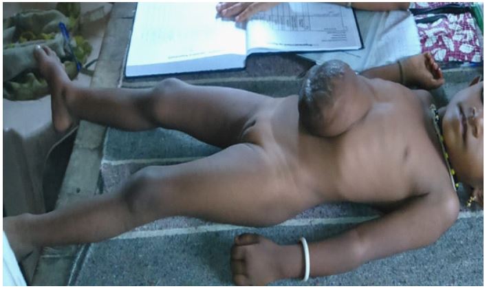
A 4-year-old girl who complained of having a huge bulging tumour at the abdomen wall presented to the OPD. On inspection, a big bag was seen to protrude through the umbilicus and outside the abdominal wall. The clinical characteristics were used to make the diagnosis of omphalocele. An uncommon congenital defect of the abdominal wall is omphalocele. Child with this giant congenital defect has covered the sac with intestinal coils, part of liver, and other abdominal organs that protrude through the umbilicus outside the abdominal wall. These organs are encased in a sac-like structure that is incredibly thin and almost translucent.
Omphalocele develops when intestinal coils fail to retract. Prenatal USG testing can be used to diagnose omphalocele in pregnant women. As a congenital anomaly, it is still present in newborns because of a lack of knowledge and inadequate prenatal screening. In this case also, quick intervention has not been started due to parents fear, poverty etc. The patient was advised to undergo surgical treatment in stages because the omphalocele was so extensive. During surgery, exposed organs are placed back into the abdomen. The abdominal opening is sealed once all the organs have been returned to the belly.
Final diagnosis: Omphalocele
Three differential diagnosis: Gastroschisis, Umbilical hernia, Physiological gut herniation.