
Journal of Clinical Images and Medical Case Reports
ISSN 2766-7820
Clinical Image - Open Access, Volume 4
Surgical emphysema and pneumothorax: Unfamiliar combination in Ehlers-Danlos syndrome
Vibha Singh; Arijit Jotdar*; Annanya Soni; Rudra Prakash; Dhruv Kapoor
Department of ENT, All India Institute of Medical Sciences, Raebareli, India.
*Corresponding Author : Arijit Jotdar
Department of ENT, Room Number 124, Hospital Block, All India Institute of Medical Sciences, Raebareli, Uttar Pradesh, India.
Tel: +91-9836896184;
Email: arijitjotdar@gmail.com
Received : Sep 21, 2023
Accepted : Oct 16, 2023
Published : Oct 23, 2023
Archived : www.jcimcr.org
Copyright : © Jotdar A (2023).
Citation: Singh V, Jotdar A, Soni A, Prakash R, Kapoor D. Surgical emphysema and pneumothorax: Unfamiliar combination in Ehlers-Danlos syndrome. J Clin Images Med Case Rep. 2023; 4(10): 2653.
Case description
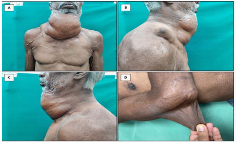
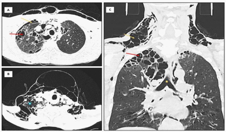
A 53-year-old gentleman presented with sudden onset swelling over the neck for the past 5 days. It was associated with dyspnoea on exertion. He was a known patient of Ehlers-Danlos syndrome since sixteen years of age. Initially, the swelling appeared over the neck which gradually progressed over to the anterior chest wall and the nape of the neck. There was no history of trauma, fever, surgery or similar complaints in the past. He was a known smoker. On examination, there was diffuse subcutaneous emphysema over the neck, and the anterior chest wall (Figure 1). Skin crepitus was present. The skin was hyper-extensile, fragile and easily stretchable (Figure 1). The patient was vitally stable. On rigid laryngoscopy, bilateral vocal cords were mobile. We admitted the patient and performed a CT scan of the neck and chest to assess the extent of the emphysema. CT scan confirmed an extensive subcutaneous emphysema over the neck, extending posterior to the oesophagus. There were multiple emphysematous bullae over the apical region of the right lung and an area of pneumothorax just in the vicinity of the bullae (Figure 2). The patient was planned for incision and evacuation of the air. A horizontal incision was given over the most prominent part of the swelling and the air was milked out gently. An Intercostal Chest Drain (ICD) was inserted and the condition was closely monitored. The recovery was uneventful. He was discharged after seven days of hospital stay.
Spontaneous pneumothorax with subcutaneous emphysema is one of the lesser-known manifestations of the Ehlers-Danlos syndrome. The pathophysiology is proposed to be the weakening of pleura and pulmonary arteries [1,2]. The lung biopsy of such patients revealed fragility of the alveolar walls that contain type III collagen. This leads to the interstitial emphysema and bleb formation [2]. The resulting respiratory manifestations can be spontaneous pneumothorax and haemothorax [3]. Due to high chance of recurrence of pneumothorax in such patients, video-associated thoracoscopic pleurodesis and application of fibrin glue is suggested.
Acknowledgement: None.
Funding: None.
Conflict of interests: We declare that we have no conflicts of interests to disclose.
Consent for publication: Written and informed consent from the patient was obtained.
References
- Dowton SB, Pincott S, Demmer L. Respiratory complications of Ehlers-Danlos syndrome type IV. Clin Genet. 1996; 50: 510-4.
- Kawabata Y, Watanabe A, Yamaguchi S, Aoshima M, Shiraki A, et al. Pleuropulmonary pathology of vascular Ehlers-Danlos syndrome: Spontaneous laceration, haematoma and fibrous nodules. Histopathology. 2010; 56: 944-50.
- Ishiguro T, Takayanagi N, Kawabata Y, Matsushima H, Yoshii Y, et al. Ehlers-Danlos syndrome with recurrent spontaneous pneumothoraces and cavitary lesion on chest X-ray as the initial complications. Intern Med. 2009; 48: 717-22.