
Journal of Clinical Images and Medical Case Reports
ISSN 2766-7820
Clinical Image - Open Access, Volume 4
CT imaging of ventriculitis: Lessons learned
*Corresponding Author : Ihssane lahlou
Laboratory of Applied Organic Chemistry, Department of Chemistry, Faculty of Sciences, University Abdelmalek Essaâdi, M’hanech II, BP 2121 Tetouan 93002, Morocco.
Email: ihssanelahlou97@gmail.com
Received : Sep 25, 2023
Accepted : Oct 17, 2023
Published : Oct 24, 2023
Archived : www.jcimcr.org
Copyright : © lahlou I (2023).
Citation: lahlou I. CT imaging of ventriculitis: Lessons learned. J Clin Images Med Case Rep. 2023; 4(10): 2655.
Description
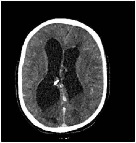
This concerns a 12-year-old female patient with a history of spina bifida for which she underwent a ventriculoperitoneal shunt at the age of 4 months. She presented to the emergency department with febrile altered consciousness associated with neck stiffness. A brain CT scan was ordered which revealed:
• Asymmetric dilation of the lateral ventricles and the 3rd ventricle, more pronounced in the right lateral ventricle, containing an isodense fluid-fluid level associated with a slight enhancement of the ventricular wall with signs of trans-ependymal resorption.
• The tip of the drain is located in the interventricular septum.
• The midline is slightly deviated to the right by 4 mm.
Discussion
Ventriculitis is an inflammation of the ependymal lining of the cerebral ventricles [1], Usually secondary to an infection. Ventriculitis can result from several causes, Including: Meningitis-bacterial and viral. Brain abscess with intraventricular rupture. Catheter-related-related to the shunt or to EVD (external ventricular drain). Trauma Cerebrospinal fluid (CSF) leakage. Complication of neurosurgery. Complications of intrathecal chemotherapy. Brain CT scans show non-specific signs notably.
• Hyperdense ventricular debris, especially in the occipital horns of the lateral ventricles,
• Hydrocephalus and periventricular low density (which probably represents reactive edema rather than transependymal edema related to hydrocephalus) are also frequently present, as may also be the features of the underlying abnormalitylow periventricular density, as well as features of the underlying anomaly (for example, signs of meningitis – abnormality or enhancement of the pial signal or of the dura/arachnoid mater)[2].
• After contrast injection, the ependymal lining of the ventricles may show homogeneous enhancement.
References
- MB Fukui, RL Williams. et al. CT and MR imaging features of pyogenic ventriculitis. AJNR Am J Neuroradiol. 2001; 22(8): 1510‑1516.
- F. Gaillard. Ventriculitis Radiology Reference Article Radiopaedia.org. Radiopaedia. 2023; https://radiopaedia.org/articles/ventriculitis.2023