
Journal of Clinical Images and Medical Case Reports
ISSN 2766-7820
Case Report - Open Access, Volume 4
Managing difficult airway in a post burn neck contracture: A case report
Manisha Sahoo; Saurabh Vig*
Department of Onco-Anaesthesia and Palliative Medicine, National Cancer Institute, Jhajjar, India.
All India Institute of Medical Sciences, New Delhi, India.
*Corresponding Author : Saurabh Vig
Department of Onco-Anaesthesia and Palliative Medicine, National Cancer Institute, Jhajjar, India. All India Institute of Medical Sciences, New Delhi, India.
Email: saurabh377@yahoo.com
Received : Sep 16, 2023
Accepted : Oct 25, 2023
Published : Nov 01, 2023
Archived : www.jcimcr.org
Copyright : © Vig S (2023).
Abstract
Post burns neck contracture is always considered a difficult airway and requires meticulous planning for proper management. Awake fiberoptic intubation (AFOI) have been gold standard for managing difficult airways [1]. Here we will discuss a case of difficult airway due to post burn contractures at the neck posted for debridement and Split Skin Grafting (SSG) for Marjolin’s ulcer at the nape of the neck. Internal changes in the airway due post inhalational injury fibrosis proved the major cause of difficulty and following traditional clinical teachings helped in successful management of airway.
Keywords: Post burn contracture; Ulcer; Burns; Neck; Deformity.
Citation: Sahoo M, Vig S. Managing difficult airway in a post burn neck contracture: A case report. J Clin Images Med Case Rep. 2023; 4(11): 2669.
Introduction
Difficult airway in post burn contractures is mainly due fibrotic bands around the nares or mouth leading to microstomia, fibrosis at the neck limiting the cervical flexion and atlantooccipital extension [2]. In addition to these external visible deformities, difficulties in airway management can arise due to unforeseen internal deformities as well. Here we describe a case of a patient with post burns contractures of face and neck posted for a procedure under general anesthesia in lateral position where difficulties in managing the airway were seen due to internal changes in the anatomy of the tracheobronchial tree.
Case report
A 39-year-old female with a childhood history of burns was posted for debridement and SSG of a non-healing Marjolijn’s ulcer at the nape of her neck. She had no other comorbidities and was a reformed tobacco chewer. On examination the patient had a fixed flexion deformity of her neck owing to mento-sternal and mandibular-clavicular contracture bands formed after burns. In addition to this fixed flexed attitude, she could not rotate her neck to either side. She had adequate oral and nasal openings. External landmarks like thyroid cartilage, cricothyroid membrane and cricoid cartilage etc. were not distinctly appreciable.
Awake fiberoptic guided oral intubation was the first plan of securing the airway, nasal intubation or emergency tracheostomy were backup plans. Patient was explained about the same during the preoperative visit and consent was taken.
On the day of surgery, induction preparation for awake fiberoptic was done with intramuscular 0.2 mg glycopyrrolate and 4% lignocaine nebulization. Inside the operation theatre she was positioned, as per her comfort because of lack of neck mobility, with sheets under her head and shoulder leaving the ulcer at the nape of the neck free (Figure 1). Standard ASA monitors were attached, airway was further prepared with viscous lignocaine gargles and xylometazoline drops to each nostril. Due to difficulty in locating the landmarks, airway blocks were avoided and spray as you go technique (SAYGO) technique with 2% lignocaine was planned. Dexmedetomidine sedation was started at 1 mcg/kg/min loading dose for 10 minutes followed by 0.5 mcg/kg/min, continuous oxygenation throughout the procedure was done with high flow nasal cannula (HFNC) with 100 percent oxygen at 50-60 liter/min.
Awake oral fiberoptic intubation was tried using ovassapian airway. The fiberoptic was passed over the tongue and through the oropharynx, uvula was seen as a small fibrosed structure, epiglottis was fixed, fibrosed, immobile and overhanging on the glottic opening which was narrowed. The fiberoptic was maneuvered with great difficulty under the overhanging fibrosed epiglottis by retroflexing and advancing beyond the overhanging edge followed by anteflexion to position the tip above the vocal cords which were sprayed with 2% lignocaine. Fiberoptic was maneuvered thorough the vocal cords into the trachea which was sprayed with 2% lignocaine. The trachea appeared narrowed and distorted. Flexometallic tube (FMT) of 7 mm was threaded over the fiberoptic but could not be negotiated beyond 10 cm. After 2 failed attempts plan of oral intubation was aborted and shifted to nasal intubation.
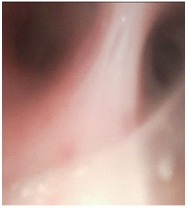
Right nostril was chosen for intubation with a 6.5 mm FMT. SAYGO technique was used again for nasal approach, airway was already anaesthetized with previous doses of local spray and trachea was approached. FMT was railroaded over the fiberoptic, and the position was confirmed visually. Patient tolerated the procedure well following which general anesthesia was induced. On ventilation chest rise appeared more on the left side and on auscultation air entry on the left side was more than right. The fiberoptic was re-inserted through the FMT and the tube position checked. The tip of FMT was found to be 3 cm above carina (Figure 2). Ventilation was resumed assuming the tube to be above carina, but airway pressures persisted to be elevated and left side air entry was more. Suspecting some anatomic anomaly, keeping in mind the distorted anatomy seen on intubation fiberoptic was reinserted and the tube was pulled back over it along with the scope to look for any anomaly in the tracheal lumen. After about 4 cm withdrawal we could see a dark opening via the murphy’s eye (Figure 3), the tube was further withdrawn revealing the carina. The tube was pulled back and fixed here following which the peak pressures settled, and air entry equalized on both the sides.
To confirm the airway anatomy fiberoptic bronchoscopy was done through the FMT, we realized that the trachea was distorted, and the right bronchus opening was steeper and anterior. On our initial fiberoptic attempts we had bypassed it directly entering the left mainstem bronchus and mistaking the left secondary carina for primary carina. The characteristic view of trachealis muscle as well as “D” shaped openings of bronchus was missing.
The case was done uneventfully, and patient was extubated inside the OT after she was fully conscious and obeying commands. She was positioned for extubation as she was for intubation. She was sent to ICU for overnight observation. She was followed up till POD2, she was pain free and had no complaints.


Discussion
Post burn contractures lead to difficult airways mainly due to hypertrophic scar tissues which restrict the range of move ments as well as the mouth opening. A proper pre anesthetic clinical evaluation of the airway is essential to formulate a proper airway management plan. There have been new additions in the field of airway management but fiberoptic guided intubation remains the gold standard for such cases [1,3]. It is the safest technique while maintaining spontaneous breaths is essential. For this a proper counselling of the patient from the preoperative day is essential.
In our case, the initial difficulty on oral fiberoptic was in negotiating tube beyond 10 cm which may have been due to the tube hinging on the fibrosed uvula. The main finding of altered anatomy and acute takeoff of the right main stem bronchus plus fibrosis and distortion of epiglottis can be attributed to the inhalational burn injury leading to inflammation and fibrosis of the airway. The mainstay of our airway management plan was oral followed by nasal fiberoptic as tracheostomy under local anesthesia would have been difficult due to absent landmarks.
The unanticipated changes in the tracheobronchial anatomy could have been assessed by a 3D CT reconstruction of the airway [4]. Such advanced techniques of airway planning have specific indications in these types of scenarios where internal anatomy may be altered.
Conclusion
In halational injury causing internal changes in the airway may pose unanticipated difficulties in securing airway in post burns patient. Having an open mind and expecting the unexpected i.e., entry of tube into the left mainstem bronchus instead of expected right sided entry in normal anatomy cases and being observant throughout the case is the key to successful airway management in such cases.
References
- Apfelbaum JL, Hagberg CA, Connis RT, Abdelmalak BB, Agarkar M, Dutton RP, et al. American Society of Anesthesiologists Practice Guidelines for Management of the Difficult Airway. Anesthesiology. 2022; 136(1): 31-81.
- Mathur R, Jain P, Chakotiya P, Rathore P. Anaesthetic and airway management of a post-burn contracture neck patient with microstomia and distorted nasal anatomy. Indian J Anaesth. 2014; 58(2): 210.
- Collins SR, Blank RS. Fiberoptic Intubation: An Overview and UpdateDiscussion. Respir Care. 2014; 59(6): 865-80.
- Calloway HE, Kimbell JS, Davis SD, Retsch-Bogart GZ, Pitkin EA, Abode K, et al. Comparison of endoscopic versus 3D CT derived airway measurements: Endoscopic Versus 3D CT Airway Measurements. The Laryngoscope. 2013; 123(9): 2136-41.