
Journal of Clinical Images and Medical Case Reports
ISSN 2766-7820
Clinical Image - Open Access, Volume 4
Melanoacanthoma simulating malignant skin lesions
Ilse Marilú Gutiérrez Villarreal1; Circe Ancona Castro2; Ely Cristina Cortés Peralta3*
1Dermatology Resident, Universidad de Monterrey, Mexico.
2Professor, Dermatology Department, Universidad de Monterrey, Mexico.
3Professor, Dermatology Department, Medical School of Tecnológico de Monterrey, Campus Monterrey, Mexico.
*Corresponding Author : Cortés Peralta EC
Professor, Dermatology Department, Medical School of Tecnológico de Monterrey, Campus Monterrey, Mexico.
Tel: +-52-81-8888 2000;
Email: dracristinacortes@tec.mx
Received : Oct 07, 2023
Accepted : Nov 02, 2023
Published : Nov 09, 2023
Archived : www.jcimcr.org
Copyright : © Cortés Peralta EC (2023).
Abstract
Cutaneous melanoacanthoma is a rare benign epithelial lesion that typically presents as a solitary black plaque or nodule in elderly people. We present a case in a 90-year-old man with a peculiar lesion on the back with conflicting clinical and dermoscopic characteristics simulating a malignant skin tumor.
Keywords: Melanoacanthoma; Malignant melanoma; Pigmented basal cell carcinoma; Pigmented lesion.
Citation: Gutiérrez Villarreal IM, Ancona Castro C, Cortés Peralta EC. Melanocanthoma simulating malignant skin lesions. J Clin Images Med Case Rep. 2023; 4(11): 2681.
Introduction
Cutaneous Melanoacanthoma (CM) is a benign epithelial tumor composed by melanocytes and keratinocytes. There are only 140 cases reported in the literature. It typically presents in individuals older than 60 years of age [1]. It is a diagnosis that has no predilection for race or sex [2]. The most common sites of presentation are head and neck, or the trunk [3]. CM presents as plaques or nodules that vary in coloration, brown or black, with an average diameter of 2 mm to 15 cm [2].
Case presentation
A 90-year-old man presented with a 40-year history of a cutaneous mass with progressive growth and signs of inflammation located on the right posterior trunk (Figure 1). Examination showed a 1 x 2 cm tumor, dark brown in color with an ulcerated center with an erythematous base, with irregular, raised, well-defined borders. Central ulceration, blue-white veil and hairpin vessels were seen on dermoscopic examination (Figure 1).
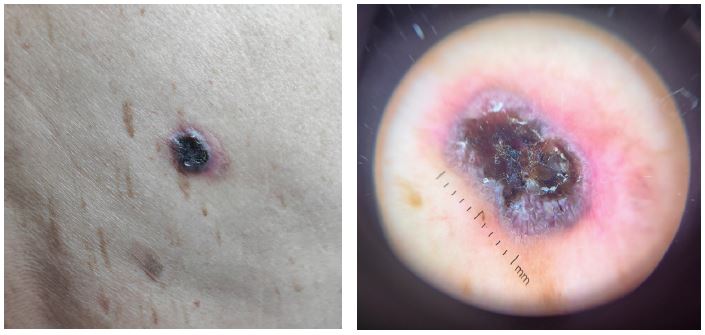

Red arrow: Blue-white veil.
Green arrow: Hairpin vessels.
Blue arrow: Erythematous base.
Discussion
The clinical differential diagnosis included malignant melanoma, pigmented basal cell carcinoma, keratoacanthoma and pigmented seborrheic keratosis. Dermoscopic examination had elements of pigmented basal cell carcinoma and keratoacanthoma. Histopathological findings were consistent with melanoacanthoma. The discrepancy between the clinical and dermoscopic characteristics in this type of lesions, in the context of a highly pigmented lesion, should be confirmed with a biopsy.
Declarations
Conflicts of interest: No conflicts of interest.
References
- Cohen Philip R, Patrick M Zito. Cutaneous Melanoacanthoma, Stat Pearls. 2023.
- Gutierrez N, Erickson CP, Calame A, Sateesh BR, Cohen PR. Melanoacanthoma Masquerading as Melanoma: Case Reports and Literature Review. Cureus. 2019; 11: e4998.
- Nakagawa K, Okabayashi A, Shimizu N, Tohda R, Imanishi H, et al. Melanoacanthoma on the genital region of a young woman. J Dermatol. 2017; 44: 729-730.