
Journal of Clinical Images and Medical Case Reports
ISSN 2766-7820
Clinical Image - Open Access, Volume 4
A rare clinical image: Papillomatosis cutis lymphostatica in a black patient
Sarah Beller1*; Disha Patel2; Billy Chacko1
1Medical College of Georgia, Augusta, Georgia, USA.
2Vascular Medicine, Harbin Clinic, Rome, Georgia USA.
*Corresponding Author : Sarah Beller
Medical College of Georgia, Augusta, Georgia, USA.
Email: sbeller@augusta.edu
Received : Sep 25, 2023
Accepted : Nov 09, 2023
Published : Nov 16, 2023
Archived : www.jcimcr.org
Copyright : © Beller S (2023).
Citation: Beller S, Patel D, Chacko B. A rare clinical image: Papillomatosis cutis lymphostatica in a black patient. J Clin Images Med Case Rep. 2023; 4(11): 2693.
Description
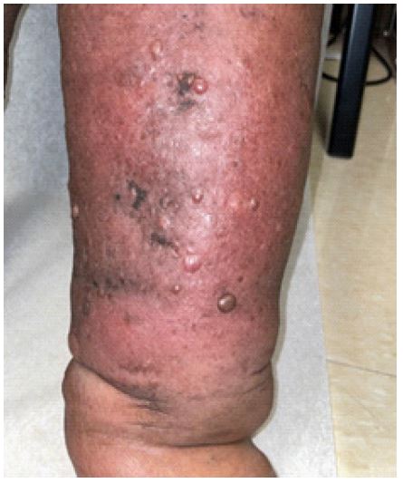
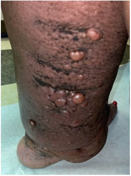
Patient is a 53 year-old black woman with a history of type 2 DM, HTN, R knee total arthroplasty, and significant obesity. She has been suffering with chronic lymphedema for several years and is presenting today with papillomatosis cutis lymphostatica with overlying cellulitis to the right lower extremity (Figures 1,2). Supporting clinical features in this patient include common features such as shiny, smooth, pearl-like, hyperkeratotic papules that are either flesh toned or hyperpigmented, positive stemmer sign, and hyperkeratotic hyperpigmented skin [1]. At this stage of pathology, edema is no longer pitting in nature. Papules range in size from 0.5 to 1 cm and are predominantly dome-shaped or occasionally scalloped (Figure 3). Today, She did have overlying erythema and discomfort upon exam due to the concurrent cellulitis, but does not typically experience pain related to the lesions. Risk factors for this patient include history of poorly controlled type [2] diabetes mellitus, right total knee replacement and varicose veins in bilateral lower extremities [1].
Key clinical message
Papillomatosis cutis lymphostatica is underrecognized in clinical settings. Access to digital visual media for clinicians is limited, articularly depicting non-caucasian patients. PCL presents potential complications such as recurrent cellulitis, disordered mobility, loss of independence and confidence, and difficulty with imaging modalities e.g. ultrasound [2].
Treatment
Mechanical, compression garments or pneumatic compression device, lymphatic massage (manual decongestion) with physical therapy [1]. Avoid mechanical measures if active infection is present, may resume once treated.
Pharmacologic: Salicylic acid ointment [3], Urea cream [3], Acitretin [4].

References
- Schultz-Ehrenburg U, Niederauer HH, Tiedjen KU. Stasis papillomatosis clinical features, etiopathogenesis and radiological findings. The Journal of Dermatologic Surgery and Oncology. 1993; 19(5): 440-446. doi:10.1111/j.1524-4725.1993. tb00371.x .
- Kerchner K, Fleischer A, Yosipovitch G. Lower extremity lymphedema update: pathophysiology, diagnosis, and treatment guidelines. J Am Acad Dermatol. 2008; 59(2): 324-31. doi: 10.1016/j.jaad.2008.04.013. PMID: 18513827.
- Gerqari A, Ferizi M, Halimi S, et al. Papillomatosis Cutis Lymphostatica - Case Report Antigona Gerqari. Journal of the Turkish Academy of Dermatology. 2016; 10.6003/jtad.16104c7.
- Feind-Koopmans A, Van de Kerkhof PC. Successful treatment of papillomatosis cutis lymphostatica with acitretin. Acta Derm Venereol. 1995; 75(5): 411. Doi:10.2340/0001555575411.