
Journal of Clinical Images and Medical Case Reports
ISSN 2766-7820
Short Report - Open Access, Volume 4
Mycosis fungoides presenting as suspected allergic reaction
John Harcha1*; Lex Leonhardt1; Keith Melvin2
1Department of Hematology Oncology Fellowship, Kettering Health, Kettering, OH, USA.
2The Jewish Hospital Department of Internal Medicine, Kenwood, OH, USA.
*Corresponding Author : John Harcha
Department of Hematology Oncology Fellowship, Kettering Health, Kettering, OH, USA.
Email: john.harcha@ketteringhealth.org
Received : Oct 24, 2023
Accepted : Nov 22, 2023
Published : Nov 29, 2023
Archived : www.jcimcr.org
Copyright : © Harcha J (2023).
Abstract
Mycosis Fungoides (MF) and Sezary Syndrome are the most common subtypes of cutaneous T cell lymphoma. MF is a mature T cell non-Hodgkin lymphoma with initial involvement of the skin, but as the disease progresses can involve lymph nodes, blood and viscera as illustrated in this case. Diagnosis is difficult due to the disease presenting similarly to benign allergic skin rashes. The median age of presentation for MF is between 50-60 years and affects males more than females. African Americans are more at risk although the exact reason is unknown.
Keywords: Mycosis fungoides; Sezary syndrome; Primary cutaneous T Cell Lymphoma; Rash.
Citation: Harcha J, Leonhardt L, Melvin K. Mycosis fungoides presenting as suspected allergic reaction. J Clin Images Med Case Rep. 2023; 4(11): 2710.
Case presentation
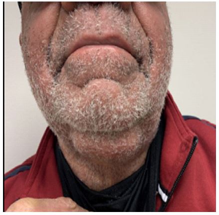
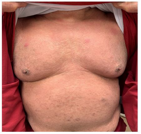
A 69-year-old African American male with history of HTN, DM and prostate cancer presented with a pruritic diffuse erythematous rash over a 6-month period. The patient has not experienced anything like this in the past nor could he identify any recent exposures or new medications. He denied any additional constitutional symptoms. On physical exam, an erythematous maculopapular rash was noted over his upper and lower extremities. Complete blood count, renal panel and immunoglobulins were unremarkable. His presentation was thought to be secondary to an allergic reaction versus drug induced reactions. He was given a Medrol dose pack and his pruritus initially improved but shortly returned following completion of his dose pack. Several of his medications were held to no relief. He was given another Medrol dose pack with the same results. He was seen by an Allergist and workup was unrevealing.
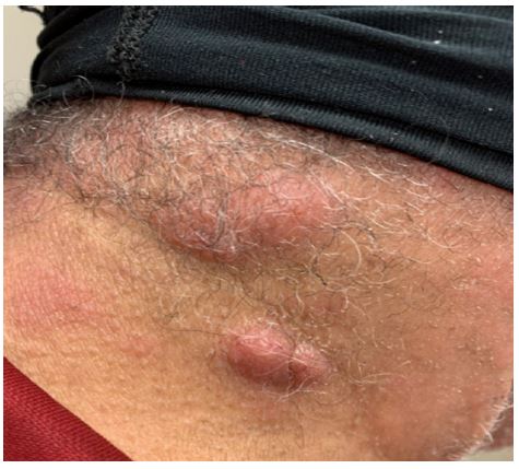
He was seen by dermatology and patient and prescribed hydrocortisone cream without much improvement. Over time, the patient’s rash seemed to progress over his entire body, now with associated facial swelling, lower extremity edema, and nodular lesions around his posterior neck and abdomen. Dermatology performed skin biopsy of posterior neck papule. Of note, he was found to have an elevated PSA and given his history of prostate CA, patient underwent PET. PET revealed multiple enlarged cervical, axillary, and inguinal lymph nodes with abnormal uptake. He was referred to Hematology/Oncology who recommended excisional lymph node biopsy of lingual LN. LN biopsy revealed atypical lymph node disorder but not diagnostic of T cell Lymphoma. Results of his skin biopsy was consistent with Mycosis fungoides. Bone marrow biopsy was consistent with T cell lymphoma and 40-50% were Sezary cells. He was started on Methotrexate 20 mg weekly and underwent extracorporeal photopheresis twice weekly. His rash and his WBC continued to improved (WBC 150k-17k). He did have a brief hospitalization for Staph bacteremia and his MTX was held and patient completed course of Ancef. MTX was resumed and unfortunately despite the improvement in his WBC and overall skin findings patient passed away of suspected cardiac etiology.
Discussion
Mycosis Fungoides (MF) and Sezary syndrome are both subtypes of cutaneous T cell lymphoma. The incidence of CTCL is 10.2 persons per million with over half of cases being MF [1]. Diagnosis is challenging given this disease process can resemble benign inflammatory or allergic dermatitis. The presentation of MF varies depending on what phase the disease process is in which includes: premycotic, patch, plaque and tumor. Diagnosis is made with skin biopsy or bone marrow biopsy for Sezary Syndrome. Staging depends on percentage of body square surface area affected, lymph node involvement, visceral organ disease, and blood involvement [2]. Treatment depends on staging and is typically skin directed in early stages and systemic in advanced stages. CTCL is typically treatable but not curable.
Conclusion
Although MF can present similarly to benign cutaneous conditions, it is an important differential to consider, especially in the African American population as there is a higher prevalence of MF. Prompt recognition and initiation of treatment is of utmost importance as it improves overall prognosis.
Declarations
Funding support: None.
Conflicts of interest statement: The authors declare that they do not have a conflict of interest.
Author contributions statement: JH, LL designed and conducted the research. KM provided the data. JH had primary responsibility for the final content. All authors read and approved the final manuscript.
References
- Jasmine Zain MD. Cutaneous T-Cell Lymphoma. Leukemia & Lymphoma Society. 2019.
- Galper SL. Diagnosis and management of mycosis fungoides. PubMed. 2010a.