
Journal of Clinical Images and Medical Case Reports
ISSN 2766-7820
Clinical Image - Open Access, Volume 4
Dermoscopy of xanthelasmoid mastocytosis
Mejjati Kaoutar*; Elloudi Sara; Douhi Zakia; Soughi Meryem; Baybay Hanane; Mernissi Fatima Zahra
Department of Dermatology, University Hospital Hassan II of Fez, Morocco.
*Corresponding Author : Mejjati Kaoutar
Department of Dermatology, University Hospital
Hassan II of Fez, Morocco.
Email: kaoutarme@hotmail.fr
Received : Oct 31, 2023
Accepted : Nov 28, 2023
Published : Dec 05, 2023
Archived : www.jcimcr.org
Copyright : © Kaoutar M (2023).
Citation: Kaoutar M, Sara E, Zakia D, Meryem S, Hanane B, et al. Dermoscopy of xanthelasmoid mastocytosis. J Clin Images Med Case Rep. 2023; 4(12): 2721.
Description
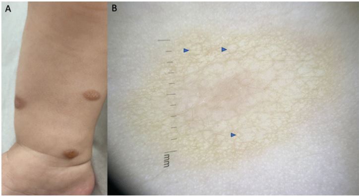
A one-year-old boy presented with a 3-months history of pruriginous cutaneous lesions on his trunk and limbs. Dermatological examination revealed multiple erythematous and pigmented plaques and nodules varying in size (Figure 1A). Darier’s sign was negative. Dermoscopy examination showed a yellowish-erythematous and homogenous background with a peau d’orange appearance. The periphery presented also pigmented stripes irradiated to the follicular openings (Figure 1B). A skin biopsy was performed, and histopathologic features were characteristic of cutaneous mastocytosis
Teaching point
Xanthelasmoid mastocytosis is a rare subtype of mastocytosis [1]. It generally occurs from birth. It presents as soft, buffyellow papules or nodules. Darier’s sign is inconsistent. Previous studies described dermoscopic patterns as brown reticular pattern, yellow-orange blot, and light-brown blot [1,2]. In our case, we found thick pigmented lines irradiated to the follicular openings distributed in a erythematous-yellowish background corresponding to melanocyte proliferation and melanin production by the high amount of mast cells. Treatment is focused on avoiding drugs and mast cell degranulation factors such as aspirin, codeine, stress, certain foods, intense physical exercise and sudden temperature changes. As far as the evolution, this clinical form is characterized by the persistence of lesions after puberty, with no increased risk of systemic damage.
References
- Chouk C, Souissi A, Raboudi A, Belkahia A, Boubaker S, Mokni M. Xanthelasmoid mastocytosis: A rare entity. Australas J Dermatol. 2021; 62: 229-230.
- Sławińska M, Kaszuba A, Lange M, Nowicki RJ, Sobjanek M, Errichetti E. Dermoscopic Features of Different Forms of Cutaneous Mastocytosis: A Systematic Review. J Clin Med. 2022; 11: 4649.