
Journal of Clinical Images and Medical Case Reports
ISSN 2766-7820
Case Report - Open Access, Volume 4
Ophthalmic shingles in children: About an unusual case
N Banane*; K Elfakiri; Gh Drais; N Rada; M Bouskraoui
Pediatric Department A, CHU Mohammed VI Marrakech Faculty of Medicine and Pharmacy, Cadi Ayyad Marrakech, Morocco.
*Corresponding Author : Najat Banane
Pediatric Department A, CHU Mohammed VI Marrakech, Faculty of Medicine and Pharmacy, Cadi
Ayyad Marrakech, Morocco.
Email: bananenajat.chu@gmail.com
Received : Nov 06, 2023
Accepted : Dec 05, 2023
Published : Dec 12, 2023
Archived : www.jcimcr.org
Copyright : © Banane N (2023).
Abstract
Shingles in children is a rare but often benign situation requiring only symptomatic treatment. The ophthalmic form is rare, which can be responsible for serious ocular complications requiring adequate and early treatment. Eye damage in children during the first four years of life is rare and potentially serious functionally if not treated in time. She must check for chickenpox in the mother during pregnancy or an area of immunosuppression.
We report the case of ophthalmic shingles in an immunocompetent 4-year-old child.
Keywords: Shingles; Child; Keratitis; Cellulite; Acyclovir.
Citation: Banane N, Elfakiri K, Drais GH, Rada N, Bouskraoui M. Ophthalmic shingles in children: About an unusual case. J Clin Images Med Case Rep. 2023; 4(12): 2731.
Introduction
Shingles is a viral dermatosis that occurs after reactivation of Varicella Zoster Virus (VZV) remaining quiescent in the dorsal sensory lymph nodes after a primary varicella infection. Its occurrence in children is exceptional, the ophthalmic form can be responsible for serious ocular complications requiring adequate and early treatment. We report the case of ophthalmic shingles in a 4-year-old child complicated by preseptal cellulitis and epithelial keratitis. After the free and informed consent of his parents.
Observation
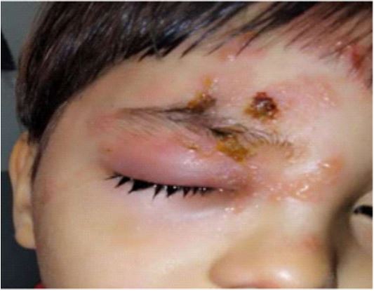
This is a 4-year-old boy, with no prior history of chickenpox, who consulted for a painful rash, affecting the forehead, the upper eyelid, the root of the nose (Figure 1).
The clinical examination revealed multiple erythematous vesicles, affecting the left hemiface, left eyelid edema, anterior segment examination: Superficial punctate keratitis at the level of the lower third of the cornea, the fundus is normal.
Craniorbital CT revealed preseptal cellulitis. The diagnosis of ophthalmic shingles was made.
A biological assessment was carried out, including fasting blood sugar and HIV serology. The results turned out to be normal.
The treatment was intravenous based on aciclovir 10 mg / kg /8 H for 7 days then relay by oral route for 2 days, antibiotic therapy based on amoxicillin clave acid, local antibiotic therapy and artificial tears were also prescribed.
The evolution is marked by the regression of the eyelid edema and the disappearance of cutaneous and ophthalmological signs.
Shingles remains rare in children and has no seasonality. The majority of cases of shingles in children occur after the age of 5. Among all reported cases of shingles, less than 10% have less than 20 years, and 5% are under 15 years of age [1], and an incidence of 0.74 per 1000 in the population under 9 years of age [1]. Shingles is always preceded by an episode of chickenpox. It is likely that the primary infection went unnoticed. The early onset of shingles can be explained by an intrauterine count of chickenpox. This observation reminds us of the possibility of occurrence of ophthalmic shingles in the pediatric population, despite the absence of a history of chickenpox episodes.
The clinical appearance of ophthalmic zoster in the acute stage is well known. It begins in 70% of cases with superficial unilateral pain at the level of the trigeminal dermatome with burning type and throbbing pain. We also note a syndrome General infectious as well as palpable pretragial lymphadenopathy. The diagnosis should be made at this stage to initiate antiviral treatment as soon as possible; 24 to 48 hours, later the erythematous, then papular, then unilateral vesicular rash appears [2].
Ocular complications occur in 50 to 70% of cases, with an often guarded prognosis [1]. They are mainly represented by keratitis, conjunctivitis, uveitis, retinitis, retinal necrosis and glaucoma. Neurological complications are possible, but fortunately rare; made of myelitis, meningoencephalitis, motor and oculomotor paralysis, bladder and digestive dysfunction. The particularity of the child’s form is the predominance of general signs, the generally favorable evolution and post-herpetic pain which remains exceptional [3,4].
Ocular complications of ophthalmic shingles require early detection and specific treatment in a specialized environment. The prescription of an antiviral must be systematic for any ophthalmic shingles. It must be started early, even if the ophthalmological examination cannot be carried out. ACYCLOVIR orally 800 mg 5x/day for 10 days if immunocompetent [5]. Intravenously: 10 mg/kg/8 hours if immunocompromised or if insufficient response to treatment started orall for 7 to 10 days. Aciclovir (Zovirax@) is used intravenously in severe forms of shingles in healthy or immunocompromised subjects, at a rate of 10 mg/ Kg/8 hours, in our observation we used the intravenous route in the face of ocular complications (epithelial keratitis).
Valaciclovir (Zelitrex@) is also indicated for the prevention of complications of ophthalmic shingles. It is a prodrug of acyclovir which has better oral bioavailability.
Acyclovir ointment can be used in combination with a systemic antiviral. Local corticosteroid therapy is indicated in immunological keratitis and anterior uveitis; General corticosteroid therapy is reserved for ophthalmic shingles complicated by acute retinal necrosis or ischemic optic neuropathy.
Associated treatments: Shower or baths once or twice a day in lukewarm water with dermatological soap without antiseptic, disinfection of skin lesions. In case of severe itching, general antihistamines can be used. In case of fever, as in chickenpox, the use of aspirin should be avoided (risk of Reye syndrome), preferring paracetamol or ibuprofen. In case of skin superinfection: oral anti-staphylococcal and anti-streptococcal antibiotic therapy.
Conclusion
Shingles is caused by reactivation of the Varicella-Zoster Virus (VZV) which remains quiescent in the dorsal sensory lymph nodes after chickenpox. The particularity of our observation is the occurrence of shingles in an immunocompetent child, and the ophthalmic localization which remains a rare form in children. Shingles ophthalmicus can cause serious eye complications. requiring adequate and early treatment. They can compromise visual function and are responsible for the severity of this condition.
References
- Infections à herpès virus de l’enfant et de l’adulte immunocompétents. La varicelle et le zona. Ann Dermatol Venereol. 2008; 135: 25-31.
- P SEMPOUX. L’atteinte oculaire dans le zona ophtalmique. LOUVAINMED. 2000; 119: 233-240.
- Banejee A. Zona de l’enfant. Archive de Pédiatrie. 1998; 5: 199- 203.
- Prise en charge des infections à VZV. Conférence de consensus de la Société de Pathologies Infectieuses de Langue Française, Lyon 25 mars 1998. Arch Pediatr. 1999; 6: 469- 76.
- Coplan P, Black S, Rojas C, Shinefeld H, Lewis E, et al. Incidence and hospitalization rates of vari- cella and herpes zoster before varicella vaccine introduction: A baseline assessment of the shifting epidemiology of varicella disease. Pediatr Inf Dis J. 2001; 20: 641-5.