
Journal of Clinical Images and Medical Case Reports
ISSN 2766-7820
Case Report - Open Access, Volume 4
Overexpressed S100A7 in the progression of oral lichen planus: From moderate dysplasia to oral squamous cell carcinoma
Mariana Gandolfo1#; Valeria Denninghoff2,3#*; Alejandra Avagnina4 ; María José Scola1 ; Pablo Turon1 ; Lidia I Adler1
1Faculty of Dentistry, University of Buenos Aires (UBA), Buenos Aires, Argentina.
2Molecular-Clinical Lab, University of Buenos Aires (UBA), National Council for Scientific and Technical Research (CONICET), Buenos Aires, Argentina.
3Liquid Biopsy and Cancer Interception Group, Centre for Genomics and Oncological Research - Pfizer – University of Granada – Andalusian Regional Government (GENyO), Granada (Andalucía), Spain
4Pathology Department, Hospital de Clínicas “José de San Martín”, Buenos Aires, Argentina.
#These authors contributed equally to this work.
*Corresponding Author : Valeria Denninghof
University of Buenos Aires (UBA), Marcelo Torcuato
de Alvear 2142, Piso 5, Sector A, C1122AAH, Buenos
Aires, Argentina.
Email: vdenninghoff@conicet.gov.ar
Received : Nov 09, 2023
Accepted : Dec 08, 2023
Published : Dec 15, 2023
Archived : www.jcimcr.org
Copyright : © Denninghof V (2023).
Abstract
The S100A7 protein plays an essential role in tumor progression and is found to be overexpressed or deregulated in oral mucosal dysplasia and several cancers. However, its expression in oral lichen planus remains unknown. Early detection of malignancy in lichen is a challenge because the histopathological diagnosis of dysplasia is subjective and is dependent on the personal assessment of each observer and there are intra and interobserver variability. In this context, using biological markers such as S100A7 could estimate its capacity for malignant transformation. The objective is to describe the immunohistochemical expression of S100A7 in a case of malignant transformation of oral lichen planus in a 62-year-old woman, in whom several foci of mild and moderate dysplasia and micro-invasive carcinoma were detected and monitored during her clinical follow-up. Immunohistochemistry of S100A7 determined increased protein expression in moderate dysplasia and micro-invasive carcinoma of oral lichen planus.
Keywords: Oral lichen planus; Malignant transformation; Early diagnosis; S100A7 protein; Immunohistochemistry.
Citation: Gandolfo M, Denninghof V, Avagnina A, Scola MJ, Turon P, et al. Overexpressed S100A7 in the progression of oral lichen planus: From moderate dysplasia to oral squamous cell carcinoma. J Clin Images Med Case Rep. 2023; 4(12): 2737.
Background
Oral lichen planus (OLP) is a chronic T-cell-mediated inflammatory disease affecting the oral mucosa and is considered one of the most frequent oral potentially malignant disorders (OPMDs) [1,2]. The concept of progressive transformation of OPMDs to oral squamous cell carcinoma (OSCC) has been well established [3]. However, it is still impossible to predict whether an OPMD will undergo malignant transformation [4,5]. In this context, oral epithelial dysplasia (OED) is one of the most reliable predictors in the assessment of the malignant capacity of OPMDs [6]. However, its histopathologic diagnosis is subjective and dependent on the assessment of each observer, generating discrepancies about which features of dysplasia are essential for predicting progression [7,8]. This situation raises the need to consider using biomarkers to estimate the capacity for malignant transformation of the oral mucosa. S100A7 is a biomarker in OPMDs and OSCC [9-12]. The S100A7 protein acts as a regulator of cell proliferation, migration, invasion, angiogenesis, and metastasis [9]. Although establishing new biomarkers to predict malignant transformation would be beneficial in managing patients with OPMDs, little is known about this antibody in lesions or precursor conditions of OSCC, particularly in OLP, and no studies on the expression of S100A7 have been reported so far.
The objective is to describe the immunohistochemical expression of S100A7 in a malignant transformation of oral lichen planus.
Case presentation
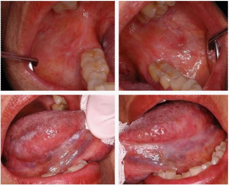
In April 2014, a 62-year-old female patient presented at the Stomatology Clinic Service of the School of Dentistry of the University of Buenos Aires, Argentina, with a tongue lesion. The patient had arterial hypertension, hypothyroidism, colon polyps, osteopenia, and OLP. During the oral cavity inspection, linear white spots were observed on both buccal mucosa at the level of the posterior thirds and an erosion in the middle third of the left buccal mucosa. On the lateral edges and ventral side of the tongue, there were white spots and keratosis or plaque (Figure 1).
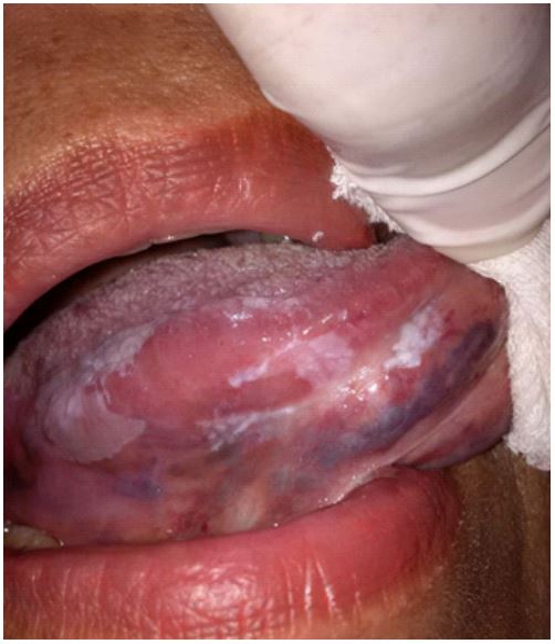
The patient was followed every six months between 2014 and 2019, a period in which different lesions appeared and were biopsied. The histological and immunohistochemical diagnosis was mild and moderate dysplasia, and therefore, they were controlled with surgical resection and local corticosteroids (Figures 2 and 3).
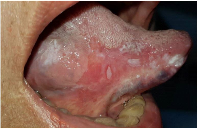
In a clinical follow-up visit in 2019, the right lateral border of the tongue had white spots, keratosis, and an ulcer surrounded by an erythematous halo (Figure 4). The patient had no lymphadenopathy.
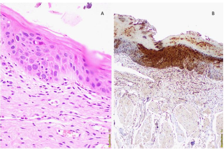
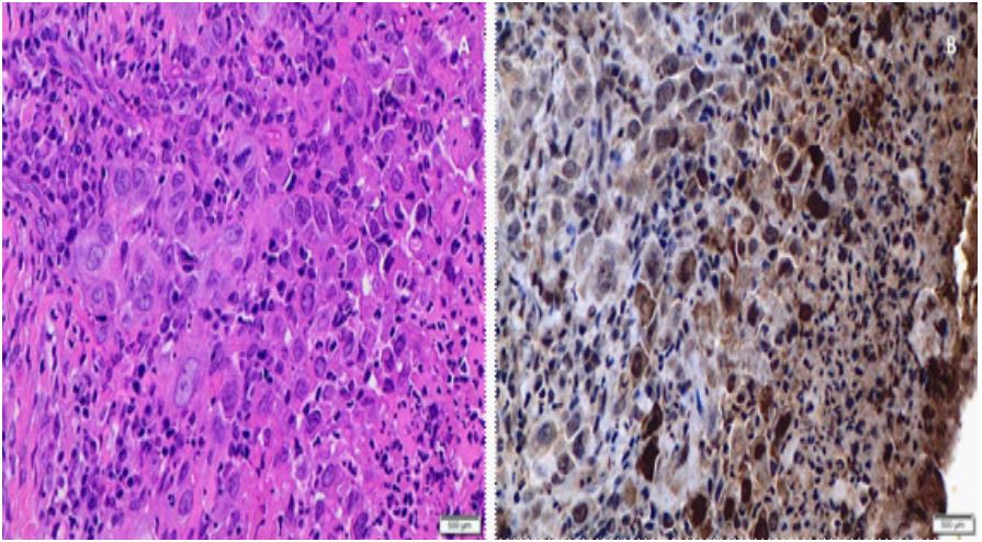
According to the characteristics of the lesion, negative lymph node palpation, buccal lichen planus, and follow-up, the presumptive clinical diagnosis was carcinoma in situ, with a differential diagnosis of OSCC in an initial or early stage. Hematological analysis and lingual ulcer biopsy for histopathological study and immunohistochemistry with S100A7 were performed (Figure 5).
The immunohistochemical study showed intense cytoplasmic and nuclear staining of S100A7 in the entire thickness of the epithelium, both in the previous lesion with moderate dysplasia illustrated in Figure 2, as in subsequent squamous cell carcinoma in Figure 5. Nuclear and cytoplasmic positivity was observed in the area with morphological alterations of dysplasia or micro-invasive carcinoma. Positivity was “patched,” with sectors where the entire thickness of the epithelium showed staining (diffuse positivity) and others where it was focal, with the positivity of some strata of the neoplastic epithelium. Histopathological features and immunohistochemical staining allowed the diagnosis of micro-infiltrating squamous cell carcinoma. The patient was referred to the María Curie Municipal Oncology Hospital for evaluation and surgical treatment and continues under strict clinical control.
Discussion
S100A7, also called Psoriasin, is a member of the S100 family of proteins initially identified as one of the most abundant proteins in psoriatic keratinocytes. It is found aberrantly expressed in malignant transformation of oral mucosal dysplasia, in some OPMD, and multiple types of cancers including OSCC, head and neck cancer, breast cancer, prostate cancer, osteosarcoma, lung cancer, ovarian cancer, and cervical cancer [13-23]. There are no papers in the literature about the value of S100A7 as a prognostic biomarker in OLP
Regarding immunohistochemical expression of S100A7 in OPMD, on the one hand, Sood et al. compared S100-A7 expression within young-onset OSCC ≤45 years (Group 1), OSCC in older age groups >45 years (Group 2), OPMD (Group 3) and inflammatory lesions (Group 4). Histological sections were classified according to the percentage of immunolabeled cells and staining intensity. Nuclear, cytoplasmic, and membrane immunoreactivity were also assessed. The OPMD group comprised mostly cases of leukoplakia, followed by erythroplakia and oral submucosal fibrosis. The authors observed that S100A7 staining (cytoplasmic and nuclear) showed a statistically significant difference between OPMD and OSCC, regardless of age. They also reported that the intensity of the reaction presented different and statistically significant intensity patterns between the OSCC group and the OPMD group. This study concluded that S100A7 can be a diagnostic biomarker to differentiate between OPMD and OSCC lesions [17]. However, the population study did not include OLP, which led us to study the expression of this protein, describing for the first time the utility of S100A7 in OLP.
In our case report, on the other hand, S100A7 is being evaluated in an ongoing clinical trial entitled “Early Prediction of Oral Cancer by S100A7 Immunohistochemistry Signature-based Assessment” (ClinicalTrials.gov ID: NCT04622462) aimed at assessing the utility of S100A7 immunohistochemical reaction to determine the risk of malignancy of clinically suspicious oral lesions. Final results are expected by December 2026 (12/31- 2026).
In our case report, the analysis of S100A7 allowed us to observe a concordance in the OLP stage between the morphology of moderate dysplasia and the expression of the protein, both at nuclear and cytoplasmic level and that in its evolution to micro infiltrating carcinoma, the intense reactivity of this protein in the tumor lesion was maintained. The immunohistochemical study performed would support the fact that the overexpression of S100A7 constitutes an early event in the malignant transformation of OLP. Considering that OSCC is the most frequent malignant neoplasm of the oral mucosa and that it is usually preceded by OPMD, early detection and early diagnosis of oral lesions at high risk of developing cancer are of utmost importance in the prevention and therapeutic approach to oral carcinogenesis in intraepithelial stages, including mild, moderate and severe dysplasia and carcinoma in situ [24,25]. In this sense, this work could be considered a hypothesis-generating work that aims to find an immunohistochemical marker to detect the progression to malignancy in OLP. The reported case highlights the malignant potential of OLP that evidences the importance of clinical follow-up and biomarkers implementation to collaborate in the early detection of malignant foci.
Conclusion
Immunohistochemical determination of S100A7 in the case of OLP presented demonstrated the expression of the protein in both moderate dysplasia and OSCC. This case report is a hypothesis generator that, if this finding is confirmed in a larger population with prolonged clinical follow-up and serial biopsies, S100A7 could be useful in the differential diagnosis between reactive lesions with reactive cellular atypia and true dysplasias.
Declarations
Conflict of interest: The authors declare no conflicts of interest.
Acknowledgement: This work was supported by a UBACyT grant (#20720170200009BA) from the University of Buenos Aires.
References
- Warnakulasuriya S. Clinical features and presentation of oral potentially malignant disorders. Oral Surg Oral Med Oral Pathol Oral Radiol. 2018; 125: 582-90. doi: 10.1016/j.oooo.2018.03.011
- Warnakulasuriya S, Kujan O, Aguirre-Urizar JM, Bagan JV, González-Moles MÁ, Kerr AR, et al. Oral potentially malignant disorders: A consensus report from an international seminar on nomenclature and classification, convened by the WHO Collaborating Centre for Oral Cancer. Oral Dis. 2021; 27: 1862-1880. doi:10.1111/odi.13704
- Tsantoulis PK, Kastrinakis NG, Tourvas AD, Laskaris G, Gorgoulis VG. Advances in the biology of oral cancer. Oral Oncol. 2007; 43: 523-534. doi:10.1016/j.oraloncology.2006.11.010
- Chang SS, Califano J. Current status of biomarkers in head and neck cancer. J Surg Oncol. 2008; 97: 640-643. doi:10.1002/jso.21023
- Warnakulasuriya S, Reibel J, Bouquot J, Dabelsteen E. Oral epithelial dysplasia classification systems: predictive value, utility, weaknesses and scope for improvement. J Oral Pathol Med. 2008; 37: 127-133. doi:10.1111/j.1600-0714.2007.00584.x
- Poh CF, Ng S, Berean KW, Williams PM, Rosin MP, Zhang L. Biopsy and histopathologic diagnosis of oral premalignant and malignant lesions. J Can Dent Assoc. 2008; 74: 283-288.
- Müller S. Oral Epithelial Dysplasia, Atypical Verrucous Lesions and Oral Potentially Malignant Disorders: Focus on Histopathology. Oral Surg Oral Med Oral Pathol and Oral Radiol. 2018; 125: 591-602. doi.org/10.1016/j.oooo.2018.02.012.
- Shubhasini A, Praveen B, Hegde U, Uma K, Shubha G, Keerthi G, et al. Inter- and Intra-Observer Variability in Diagnosis of Oral Dysplasia. Asian Pac J Cancer Prev. 2017; 18: 3251-3254. doi:10.22034/APJCP.2017.18.12.3251
- Nikitakis NG, Pentenero M, Georgaki M, Poh CF, Peterson DE, Edwards P, et al. Molecular markers associated with development and progression of potentially premalignant oral epithelial lesions: Current knowledge and future implications. Oral Surg Oral Med Oral Pathol Oral Radiol. 2018; 125: 650-669. doi:10.1016/j.oooo.2018.03.012
- Raffat MA, Hadi NI, Hosein M, Mirza S, Ikram S, Akram Z. S100 proteins in oral squamous cell carcinoma. Clin Chim Acta. 2018; 480: 143-149. doi:10.1016/j.cca.2018.02.013
- Probstmeier R, Kraus D, Wenghoefer M, Winter J. S100 Proteins as Biomarkers in Risk Estimations for Malignant Transformation in Oral Lesions. Methods Mol Biol. 2019; 1929: 763-771. doi:10.1007/978-1-4939-9030-6_48.
- Sivadasan P, Gupta MK, Sathe G, Sudheendra HV, Sunny SP, Renu D, et al. Salivary proteins from dysplastic leukoplakia and oral squamous cell carcinoma and their potential for early detection. J Proteomics. 2020; 212: 103574. doi:10.1016/j.jprot.2019.103574
- Kataoka K, Ono T, Murata H, Morishita M, Yamamoto KI, Sakaguchi M, et al. S100A7 promotes the migration and invasion of osteosarcoma cells via the receptor for advanced glycation end products. Oncol Lett. 2012; 3: 1149-1153. doi:10.3892/ol.2012.612
- Raffat MA, Hadi NI, Hosein M, Mirza S, Ikram S, Akram Z. S100 proteins in oral squamous cell carcinoma. Clin Chim Acta. 2018; 480: 143-149. doi:10.1016/j.cca.2018.02.013.
- Liu G, Wu Q, Liu G, Song X, Zhang J. Knockdown of S100A7 reduces lung squamous cell carcinoma cell growth in vitro and in vivo. Int J Clin Exp Pathol. 2014; 7: 8279-8289.
- Kaur J, Matta A, Kak I, Srivastava G, Assi J, Leong I, et al. S100A7 overexpression is a predictive marker for high risk of malignant transformation in oral dysplasia. Int J Cancer. 2014; 134: 1379-1388. doi:10.1002/ijc.28473.
- Sood A, Mishra D, Kharbanda OP, Chauhan SS, Gupta SD, Deo SSV, et al. Role of S100 A7 as a diagnostic biomarker in oral potentially malignant disorders and oral cancer. J Oral Maxillofac Pathol. 2022; 26: 166-172. doi:10.4103/jomfp.jomfp_402_20.
- Gagnon A, Kim JH, Schorge JO, Ye B, Liu B, Hasselblatt K, et al. Use of a combination of approaches to identify and validate relevant tumor-associated antigens and their corresponding autoantibodies in ovarian cancer patients. Clin Cancer Res. 2008; 14: 764-771. doi:10.1158/1078-0432.CCR-07-0856
- Wang R, Li Y, Hu E, Kong F, Wang J, Liu J, et al. S100A7 promotes lung adenocarcinoma to squamous carcinoma transdifferentiation, and its expression is differentially regulated by the HippoYAP pathway in lung cancer cells. Oncotarget. 2017; 8: 24804-24814. doi:10.18632/oncotarget.15063
- Wang R, Li Y, Hu E, Kong F, Wang J, Liu J, et al. S100A7 promotes lung adenocarcinoma to squamous carcinoma transdifferentiation, and its expression is differentially regulated by the HippoYAP pathway in lung cancer cells. Oncotarget. 2017; 8: 24804-24814. doi:10.18632/oncotarget.15063
- Lu Z, Zheng S, Liu C, Wang X, Zhang G, Wang F, et al. S100A7 as a potential diagnostic and prognostic biomarker of esophageal squamous cell carcinoma promotes M2 macrophage infiltration and angiogenesis. Clin Transl Med. 2021; 11: e459. doi:10.1002/ctm2.459
- Nasser MW, Wani NA, Ahirwar DK, Powell CA, Ravi J, Elbaz M, et al. RAGE mediates S100A7-induced breast cancer growth and metastasis by modulating the tumor microenvironment. Cancer Res. 2015; 75: 974-985. doi:10.1158/0008-5472.CAN-14-2161
- Nasser MW, Qamri Z, Deol YS, Ravi J, Powell CA, Trikha P, et al. S100A7 enhances mammary tumorigenesis through upregulation of inflammatory pathways. Cancer Res. 2012; 72: 604-615. doi:10.1158/0008-5472.CAN-11-0669.
- Lambert R, Sauvaget C, de Camargo Cancela M, Sankaranarayanan R. Epidemiology of cancer from the oral cavity and oropharynx. Eur J Gastroenterol Hepatol. 2011; 23: 633-641. doi:10.1097/MEG.0b013e3283484795.
- Warnakulasuriya S, Johnson NW, van der Waal I. Nomenclature and classification of potentially malignant disorders of the oral mucosa. J Oral Pathol Med. 2007; 36: 575-580. doi:10.1111/j.1600-0714.2007.00582.x