
Journal of Clinical Images and Medical Case Reports
ISSN 2766-7820
Case Report - Open Access, Volume 4
Human urogenital myiasis caused by Clogmia albipunctata (Diptera: Psychodidae): First report in Bangladesh
Ariful Bashar1; Rupen Nath2; Proggananda Nath3; Debashis Ghosh2; Rajib Chowdhury2,4*
1Infectious Diseases Hospital, Mohakhali, Dhaka-1212, Bangladesh.
2International Centre for Diarrhoeal Disease Research, Bangladesh (ICDDR,B), Dhaka-1212, Bangladesh.
3Mymensingh Medical College and Hospital (MMCH), Mymensingh-2200, Bangladesh.
4Independent University Bangladesh (IUB), Dhaka-1229, Bangladesh.
*Corresponding Author : Rajib Chowdhury
International Centre for Diarrhoeal Disease Research, Bangladesh (ICDDR,B), Dhaka-1212, Bangladesh.
Email: rajib478@yahoo.com
Received : Nov 12, 2023
Accepted : Dec 11, 2023
Published : Dec 18, 2023
Archived : www.jcimcr.org
Copyright : © Chowdhury R (2023).
Abstract
We report the first case of human urogenital myiasis caused by Clogmia albipunctata in a 32-year-old housewife in Bangladesh. Identification at the species level has been performed by morphological examination of exerted pupa and adult flies generated from pupa by rearing in the entomology laboratory, and they belonged to Clogmia albipunctata. Ivermectin was administered to the patient as medication, and finally, all larvae and pupae were completely gone from the urine. Assumedly, this is the first description of human urogenital myiasis in Bangladesh, and it will also shed some light on the medical importance and management of urinary myiasis.
Keywords: Clogmia albipunctata; Urogenital myiasis; Bangladesh.
Citation: Bashar A, Nath R, Nath P, Ghosh D, Chowdhury R. Human urogenital myiasis caused by Clogmia albipunctata (Diptera: Psychodidae): First report in Bangladesh. J Clin Images Med Case Rep. 2023; 4(12): 2739.
Introduction
Urogenital myiasis is a rare physical condition that is seen in immunocompromised individuals, the elderly, and people with poor personal hygiene. It commonly occurs in tropical and subtropical countries and areas with warm climates [1,2]. In 1840, “myiasis”, the term first introduced by Hope, was derived from the Greek word “myia”, which means ‘fly’, and that describes the infestation of tissues or organs of vertebrate animals, domestic or wild, and humans [3]. The dipteran (two-winged) fly species maggots feed to survive in the host’s tissue, body cavity, and liquid body substances at least for a certain period [3,4] and could cause a broad range of infestations, depending on the body location and the relationship of the larvae with the host [5]. Larvae, as well as maggots, can enter not only through skin wounds or body cavities (mouth, ears, eyes, and urogenital tract) but also be able to pierce and penetrate both healthy and necrotic tissues, which originate a secondary infection as complications of myiasis [6].
The three types of myiasis-obligatory, facultative, and accidental-are classified based on the relationship between the host and larvae [7,8]. The obligatory myiasis refers to the cause of fly larvae that obligately require host living tissue to survive; the facultative myiasis is the cause of those fly larvae that infest host wounded or necrosing tissue to survive; and those fly larvae that are accidentally ingested and accumulate on tissues of humans or animals may be responsible for accidental myiasis [8]. Moreover, human myiasis is classified based on the part or organ of the body involved and on the relationship between the host and larvae. The involvement of body organs can present as cutaneous (dermal or sub-dermal) myiasis, anal myiasis, nasopharyngeal myiasis, ocular myiasis, body cavity myiasis, wound myiasis, ophthalmic myiasis, aural myiasis, gastrointestinal myiasis, and Urogenital Myiasis (UGM) [1,7].
Myiasis occurs worldwide, with more cases being reported from tropical, subtropical, and warm temperate areas. Bangladesh throws some light on the medical importance and management of this disease in our area. In our presentation, we aimed to present a case of urogenital myiasis caused by the Clogmia albipunctata fly.
Case presentation
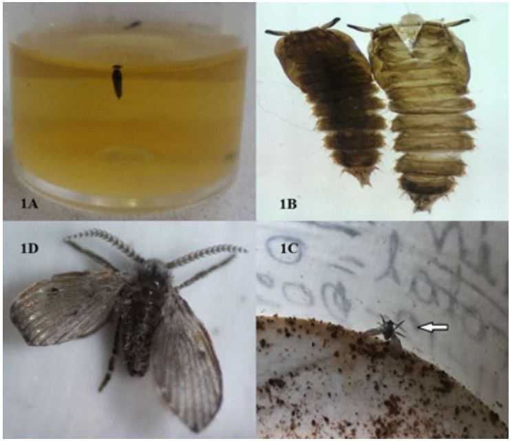
A 32-year-old woman came to the Outpatient Department (OPD) of Surya Kanta Hospital (SK-hospital) in Mymensingh, Bangladesh, with a half glass of highly colored water containing various black elements floating inside. She lived in Dhobaura upazila, in the northern region of the Mymensingh Division of Bangladesh. Her complaint was an aching feeling all over the body, discomfort, and painful urination. She claimed that the glass containing urine and the regularly observed small insectlike objects passed her urine at different times over one year. She looks panicked, restless, and frustrated, as proven by her complaints. She did urinate within the hospital lavatory and collected the same type of urine (Figure 1A). The patient lives in a remote area and maintains personal hygiene, but took a regular bath in the pond. Her socioeconomic condition is average; she lives in a semi-paka house and obtains drinking water from a tube well.
Informed consent was taken from the patient, after explaining the whole procedure to her.
The patient had no previous history of significant medical or surgical history except for a right-sided laparoscopic ovarian cystectomy about 5 years ago due to single large and small multiple cysts. During the physical examination, no abnormalities were found, and the doctor recommended some laboratory investigations, including a routine urine examination, a gram stain, and a culture with sensitivity (C&S). All investigations revealed no abnormalities in patients except for a urine stain that showed larvae or pupae of an unknown parasite (insect). In these circumstances, the patient was asked to collect up to 24 hours’ worth of urine samples to confirm the information claimed by the patient. Over the following hours, 4 small, grayish-black-colored warms were examined under the microscope, and they were diagnosed as pupae of a suspected Clogmia species (Figure 1A,1B), which revealed the authenticity of the claim by the patient. The pupae were actively motile, and pyriform in shape, with a cephalothorax carrying two antennae and a segmented abdomen. Their size ranged from 4–5 mm in length. One pupa was transferred to a plastic pot with humid plaster of Paris and was taken to the insectarium incubator, maintaining a 12:12 (day: night) photoperiod at 27°C -28°C temperature with a relative humidity of 75% at the entomology unit in SK hospital, which was being managed by the International Centre for Diarrhoeal Disease Research, Bangladesh (icddr,b). After three days, a newly emerged fly (Figure 1C) was found inside that pupa. The fly’s identification was microscopically confirmed as Clogmia albipunctata (Figure 1D) according to the external morphological characteristics of its wings, which are gray or brown and hairy (setae); antennae with dense setae, each segment with distinctive whorls; pointed wings with some white spots; and usually held parallel to the substrate when at rest. Finally, it was diagnosed that the patient had ‘urogenital myiasis’ a very rare case in Bangladesh. The patients were recommended to take Ivermectin in a single oral dose of 200 µg/Kg for 2 months. In addition, antibiotics and antiseptic therapy were given to prevent secondary bacterial infection, and the patient was asked to take plenty of oral hydration to drive away any larval or pupal materials in the bladder. A complete recovery of symptoms was observed at the end of the post-therapeutic follow-up.
Discussion
Urogenital myiasis in humans is a very rare and uncommon clinical condition in several countries in the world [9]. It is to be found almost exclusively in females than in males, and it is assumed, perhaps due to the physiological and anatomical features of the urogenital system and poor hygiene practices [2]. The family Psychodidae is subdivided into six subfamilies, only two of which have medical importance for humans: Phlebotominae (sandflies), which are bloodsuckers and vectors of leishmaniasis, and Psychodinae (moth flies), which are not adapted for bloodsucking [10]. Some Psychodinae and Dipteran species are responsible for originating urogenital myiasis, including Psychoda albipennis [11,12], Lucilia sericata, Whlfahartia magnifica [13], Fannia canicularis [14], and Clogmia albipunctata [15]. The way reports of human urogenital myiasis are noticed in different parts of the world is not found in Bangladesh.
To our knowledge, this is the first case report of human urinary myiasis caused by C. albipunctata in Bangladesh. Unfortunately, this patient suffered from urogenital myiasis with dysuria, a mild fever, and itching in the peritoneal region for a long time (more than one year) due to its misdiagnosis. Even after repeated urine tests had been done in several laboratories, however, they failed to accomplish a correct diagnosis of urogenital myiasis. From then on, she was treated for those symptoms but not cured. Most species of Psychodinae (moth flies) breed in bathrooms, toilets, drains, drain pipes, or septic tanks, and due to their non-biting nature, they are known as domestic insects to local people [15,16]. It was previously suggested that flies lay their eggs with poor personal hygiene when urinating in or around the moist urogenital orifices, which is the suitable environmental region for hatching eggs. After hatching, larvae climb to the urinary tract to feed in order to survive, which is infested by myiasis and is estimated to be present in our patient [10,13]. In this case, the finding of an infestive, authentic immotile pupa and destroyed body parts of larvae served as diagnostic evidence. The diagnosis of true urinary myiasis was confirmed through repeated emissions of pupa with destroyed parts of the larva; moreover, the new adult flies from the excreted pupae were colonized for 3 days in the entomology laboratory.
For removing larvae or pupae, Ivermectin is used as the main drug for treating myiasis in humans or animals, and then to prevent secondary infections, systemic treatment with broadspectrum antibiotics is applied. Surgical excision may also be an alternative option to removal, although it is not always necessary [1]. The patient was treated with Ivermectin as a drug and plentiful oral hydration. As a result, the patient completely recovered from her symptoms.
Conclusion
Various adult moth flies are normally found in a dark, moist region in the local area, but people are not concerned about them due to a lack of awareness regarding their clinical importance as well as their belief that they are only nuisance pests. This point of view should be changed to decrease the manifestation of urinary myiasis. This detected case of urinary myiasis in Bangladesh will draw attention from health policymakers to the rising awareness of the medical importance and management of this disease. In addition, it also draws the attention of urologists and laboratorians towards the possibility of detecting larvae, pupae, or their fragments in the urine of dipteran flies. So that they do not avoid these things as contaminants and keep in mind that they can lead to an abnormal cause of urinary tract infection.
Declarations
Acknowledgment: We would like to thank the patient for participating in the study. We are so grateful to the entomological technicians at the Entomology Unit of SK Hospital for their help in rearing the Clogmia albipunctata flies in the laboratory.
Funding source: The authors have not received any specific funds to conduct the present study.
Competing interests: None declared.
References
- Sapre AS, Natu VN, Patel MV, Chandwaskar N. Rare Case of Urogenital Myiasis. J Obstet Gynecol India. 2013; 63: 145-146.
- Singh A, Kaur J. Occurrence of human urogenital myiasis due to neglected personal hygiene: A review. Trans R Soc Trop Med Hyg. 2019; 113: 4-10.
- Mahmud S, Islam M, Rahman M, Alam M, Rahman M, Litu M. Myiasis in a the Tracheotomy Wound: A Case Report. Bangladesh J of Otorhinolaryngol. 2020; 22: 119-121.
- Culha M, Turker K, Ozsoy S, Serefoglu E. Urogenital myiasis caused by Psychoda albipennis. Saudi Med J. 2016; 37: 1392-1394.
- Myiasis. Clin Microbiol Rev. 2020.
- Faridnia R, Soosaraei M, Kalani H, Fakhar M, Jokelainen P, et al. Human urogenital myiasis: A systematic review of reported cases from 1975 to 2017. Eur J Obstet Gynecol Reprod Biol. 2019; 235: 57-61.
- Akinci O, Sirekbasan S, Toksoy M, Ergun S. A Case of Breast Myiasis Caused by Lucilia cuprina. Am J Med Case Rep. 2017; 5: 196-198.
- Ramana KV. Human Myiasis. J Med Microbiol Diagn. 2012.
- Gashout A, Amro A, Hamarsheh O, Al-Dwibe H. Urogenital Myiasis Caused by Psychoda albipennis in a Female Child in Libya. Turk J Parasitol. 2019; 43: 152-154.
- El-Dib NA, Wahab WMAE, Hamdy DA, Ali MI. Case Report of Human Urinary Myiasis Caused by Clogmia albipunctata (Diptera: Psychodidae) with Morphological Description of Larva and Pupa. J Arthropod Borne Dis. 2017; 11: 533-538.
- Yenice MG, Demir T, Babür C, Nalbantoğlu S, Kılıç S. A case of urogenital myiasis caused by Psychoda albipennis (Diptera: Nematocera). Mikrobiyol Bul. 2011; 45: 558-564.
- Oguz U, Resorlu B, Cizmeci Z, Unsal A. A rare urogenital myiasis caused by Psychoda albipennis: A case report. Turk J Urol. 2012; 38: 168-169.
- Salimi M, Goodarzi D, Karimfar M, Edalat H. Human Urogenital Myiasis Caused by Lucilia sericata (Diptera: Calliphoridae) and Wohlfahrtia magnifica (Diptera: Sarcophagidae) in Markazi Province of Iran. Iran J Arthropod Borne Dis. 2010; 4: 72-76.
- Perez-Eid C, Mouffok N. Human urinary myiasis caused by Fannia canicularis (Diptera, Muscidae) larvae in Algeria. Presse Med. 1999; 28: 580-581.
- Hjaija D, Sawalha S, Amr Z, Katbeh-Bader A, Hassoon R. Urinary Myiasis Caused by Clogmia albipunctata from the Palestinian Territories. Bull Soc Pathol Exot. 2018; 111: 148-151.
- Kvifte GM, Wagner R. 24 PSYCHODIDAE (Sand Flies, Moth Flies or Owl Flies), Manual of Afrotropical Diptera, Nematocerous Diptera and lower Brachycera. Suricata 5. South African National Biodiversity Institute, Pretoria; 2017: 2: 607-632.