
Journal of Clinical Images and Medical Case Reports
ISSN 2766-7820
Clinical Image - Open Access, Volume 4
Neurosarcoidosis; the great mimicker
Shamim Kazemi1; Minoo Rouhi1 *; Mahsa Sepahvand1; Mohammad Rohani1; Narges Yazdi2
1Department of Neurology, Rasool Akram Hospital, School of Medicine, Iran University of Medical Sciences, Tehran, Iran.
2Department of Neurology, School of Medicine, Iran University of Medical Sciences, Tehran, Iran.
*Corresponding Author : Minoo Rouhi
Department of Neurology, Rasool Akram hospital,
School of Medicine, Iran University of Medical Sciences, Tehran, Iran.
Tel: 09112431937
Email: minoorhi72@gmail.com
Received : Nov 18, 2023
Accepted : Dec 13, 2023
Published : Dec 20, 2023
Archived : www.jcimcr.org
Copyright : © Rouhi M (2023).
Abstract
We report a 36 Y/O male that present with seizure and a solitary mass-like lesion in brain MRI which was proven to be a non-caseating granulomatous inflammation by biopsy in the context of Neurosarcoidosis.
Keywords: Neurosarcoidosis; Brain mass; Non-caseating granulomatous inflammation.
Citation: Rouhi M, Kazemi S, Sepahvand M, Rohani M, Yazdi N. Neurosarcoidosis; the great mimicker. J Clin Images Med Case Rep. 2023; 4(12): 2745.
Description
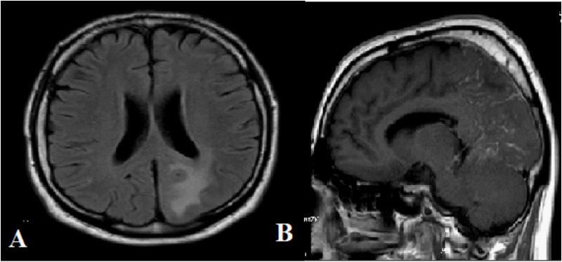
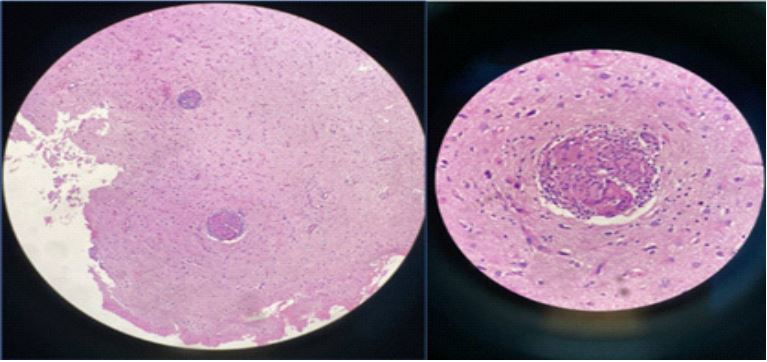
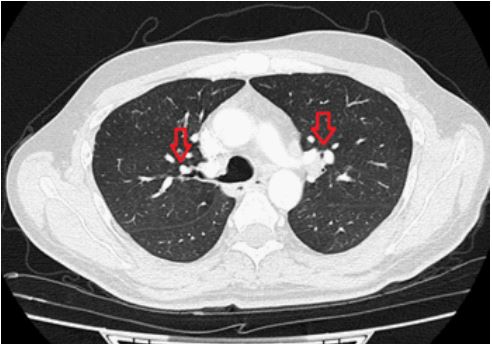
The patient was a 35 years old man who presented with focal onset seizure with impaired awareness, headache and left hemianopia in the past 3 month. Brain MRI (Magnetic Resonance Imaging) revealed abnormal low T1/high T2 signal intensity with focal nodular leptomeningeal enhancement in periventricular, deep and subcortical white matter of left high parieto-occipital without restriction (Figure 1). The brain biopsy showed non-caseating granulomatous inflammation. Ziehl-Neelsen staining for acid-fast Bacillus was negative (Figure 2). The spiral Chest computed topography (CT) scan taken in 2 months follow up included bilateral symmetric hilar lymphadenopathy (Figure 3). Serum ACE level was 59 U/L (8-52 U/L) but normal CSF (Cerebrospinal fluid) level. He was diagnosed with Neurosarcoidosis and after 5 gram of methylprednisolone injection, Infliximab (the TNF alpha inhibitor agent) started for him. Intracranial granulomatous mass lesions can present in 5-10% of Neurosarcoidosis patients as a tumefactive mass, mimicking a tumor [1-5] .
References
- Bradshaw, M.J., et al., Neurosarcoidosis: pathophysiology, diagnosis, and treatment. Neurology-Neuroimmunology Neuroinflammation. 2021; 8(6).
- Ungprasert, P. and E.L. Matteson, Neurosarcoidosis. Rheumatic Disease Clinics. 2017; 43(4): 593-606.
- Ginat, D.T., G. Dhillon, and J. Almast, Magnetic resonance imaging of neurosarcoidosis. Journal of Clinical Imaging Science, 2011; 1: 15.
- Bathla, G., et al., Imaging of neurosarcoidosis: common, uncommon, and rare. Clinical radiology. 2016. 71(1): 96-106.
- Lury, K.M., et al. Neurosarcoidosis--review of imaging findings. in Seminars in roentgenology. 2004; 39(4): 495-504.