
Journal of Clinical Images and Medical Case Reports
ISSN 2766-7820
Clinical Image - Open Access, Volume 4
Delayed onset Nevus of Ota: A rare presentation in a 60-year-old woman
*Corresponding Author : Muhammad Arif Ozir
Department of Ophthalmology, Hospital Pengajar
Universiti Sultan Zainal Abidin, Terengganu,
Malaysia.
Email: drmuhammadarif@yahoo.com
Received : Nov 18, 2023
Accepted : Dec 13, 2023
Published : Dec 20, 2023
Archived : www.jcimcr.org
Copyright : © Arif Ozir M (2023).
Abstract
Nevus of Ota is usually characterized by unilateral, mottled, slate blue or dark brown hyperpigmentation on the forehead, face and around eye area. Ota’s nevus is usually congenital but may appear in early childhood or in puberty. We report a late onset Nevus of Ota in a 60-year-old Asian woman and discuss clinical diagnosis, risks, and treatment modalities.
Keywords: Nevus of Ota; Oculodermal melanocytosis; Glaucoma; Melanoma.
Citation: Arif Ozir M. Delayed onset Nevus of Ota: A rare presentation in a 60-year-old woman. J Clin Images Med Case Rep. 2023; 4(12): 2747.
Description
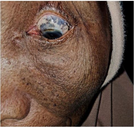
A 60-year-old Asian woman presented with left eye hyperpigmentation during a mobile ophthalmology medical checkup at rural village area. The hyperpigmentation is left ipsilateral and involves the region of forehead, cheeks, and side of the face. Upon further history, she insisted that the hyperpigmentation had been presented only since one year ago and become darker in color since then. She has no other ocular complaints such as redness, pain, tearing, vision loss, flashes, or floaters. There is no intraoral hyperpigmentation. During the presentation, the vision is 20/20. The oblique flashlight test is normal and digitally normotensive for intraocular pressure estimation. Anterior segment examination shows scleral and eyelid hyperpigmentation (Figure 1). Confrontation visual field examination is normal. Fundus examination optic nerve shows no sign of glaucomatous disc with a 0.3 cup-to-disc ratio and retina examination shows no sign of melanoma. She was offered to be referred to our eye center for further follow-up and dermatology referral for treatment. She denies the follow-up offer and treatment and wants to be conservative since it did not affect her vision.
Discussion
Oculodermal melanosis (Nevus of Ota, melanosis oculi) is a benign mesodermal melanosis involving the distributions of the ophthalmic and maxillary trigeminal nerve with associated hyperpigmentation of the eye and its adnexa [1]. Entrapment of melanocytes in the upper third of the dermis leads to gray-blue macular hyperpigmentation of the conjunctiva and sclera and ipsilateral facial skin, usually occurring unilaterally [2]. Nevus of Ota typically presents at birth but can also appear in puberty or during pregnancy [3]. Isolated cases of delayed onset acquired Nevus of Ota that first appear in adults, including in older patients, have been reported although it is rarer [4]. Nevus of Ota presents more commonly in females than males, with a 5:1 ratio [5]. It also occurs predominantly in people of Asian and African descent [6].
Patients with Nevus of Ota are at risk for glaucoma and melanoma [5]. Those with Nevus of Ota extending into the eye have an increased risk of developing glaucoma (10% of patients) as invasion of melanocytes can block drainage of aqueous, leading to elevated intraocular pressures [7]. 1/400 patients can develop uveal melanoma (typically choroidal) in the affected eye. Risk factors for malignant transformation include cutaneous or palatal melanocytosis, scleral involvement of the superior, nasal, or temporal quadrants, choroidal melanocytosis and diffuse iris melanocytosis [8,9].
The most effective treatment approach for cosmetic treatment of Nevus of Ota is via laser therapy. Treatment is usually not required unless for malignant transformation of skin lesions or for cosmetic reasons.
Nevus of Ota is typically benign with excellent ophthalmology and dermatology prognosis. Screening for glaucoma and malignant melanoma by an ophthalmologist and dermatologist is recommended yearly [5]. In this case, the patient denies any further intervention in view of still having good vision.
Declarations
Ethical approval: Not required.
Declaration of conflicting interests: None declared.
Funding: No funding sources.
References
- Rapini RP, Bolognia JL, Jorizzo JL. (2007). Dermatology: 2-Volume Set. St. Louis: Mosby. pp. 1720 22.
- Bhattacharya, S. K., Girgla, H. S., & Singh, G. (1973). Nevus of Ota. International Journal of Dermatology, 12(6), 344-347.
- Kopf, A. W., & Weidman, A. I. (1962). Nevus of ota. Archives of Dermatology, 85(2), 195-208.
- Singh AP, Bagewadi A, Keluskar V, Shetti A. Nevus of Ota involving palate: case reports and review. Journal of Indian Academy of Oral Medicine and Radiology. 2007;19:441–45. [Google Scholar].
- Cronemberger, S., Calixto, N., & Freitas, H. L. (2011). Nevus of Ota: clinical-ophthalmological findings. Revista Brasileira de Oftalmologia, 70(5), 278-283.
- Shetty SR, Subhash BG, Rao KA, Castellino R. Nevus of Ota with buccal mucosal pigmentation. Dent Resj (Isfahan) 2011 Winter, 8 (11) 52-5.
- Shaffer, D., Walker, K., & Weiss, G. R. (1992). Malignant melanoma in a Hispanic male with nevus of Ota. Dermatology, 185(2), 146-150.
- Redkar, N. N., Rawat, K. J., Warrier, S., & Jena, A. (2016). Nevus of Ota. Journal of the Association of Physicians of India, 64, 70.
- Hannan, C. (2018). Nevus of Ota: Treatment, Eye, Removal, Glaucoma, and More. Healthline. Retrieved at: https://www.healthline.com/health/nevus-of-ota