
Journal of Clinical Images and Medical Case Reports
ISSN 2766-7820
Clinical Image - Open Access, Volume 4
Ectropion in a collodion baby: A rare case report
Swatishree Nayak, MS, FPOS1*; Shalvika Gupta, MBBS2 ; Chinmaya Kumar Panda, MD, PDCC (Critical Care)3
1Assistant Professor, Department of Ophthalmology, All India Institute of Medical Sciences, Raipur, India.
2Junior Resident, Department of Ophthalmology, All India Institute of Medical Sciences, Raipur, India.
3Associate Professor, Department of Anaesthesiology and Critical Care, All India Institute of Medical Sciences, Raipur, India.
*Corresponding Author : Swatishree Nayak
Assistant Professor, Department of Ophthalmology,
All India Institute of Medical Sciences, Raipur, India.
Email: nswatishree@yahoo.com
Received : Nov 20, 2023
Accepted : Dec 14, 2023
Published : Dec 21, 2023
Archived : www.jcimcr.org
Copyright : © Nayak S (2023).
Abstract
Collodion baby is a rare congenital disorder characterized by parchment like tight membrane covering the whole body. This extra layer of skin with its compressive effects can lead to cicatricial lid changes, ectropion and secondary corneal exposure. We report a case of a twenty-four day old collodion baby who presented with bilateral eyelid ectropion that was timely managed with local ocular treatment and systemic acitretin. The management of collodion baby is challenging, but early diagnosis and interdisciplinary involvement can optimize outcomes and improves prognosis.
Keywords: Collodion baby; Cicatricial; Ectropion.
Citation: Nayak S, Gupta S, Panda CK. Ectropion in a collodion baby: A rare case report. J Clin Images Med Case Rep. 2023; 4(12): 2749.
Introduction
Collodion baby is a term used for neonates who are born with a thick translucent parchment like sheet encasing the whole body [1]. This membrane is a result of epidermal hyperkeratosis due to dysfunctional epidermal development. This rare condition has an incidence of 1 in 50,000 -100,000 deliveries [2]. Despite the known complications associated with this condition, these neonates should be provided with basic supportive neonatal care promptly to improve prognosis. These new-borns are especially susceptible to exposure keratopathy as a sequela to congenital ectropion which may further progress to sight threatening infectious keratitis [3]. We report a case of a collodion baby with bilateral eyelid ectropion that showed signs of improvement on receiving local ocular treatment and systemic acitretin.
Case report
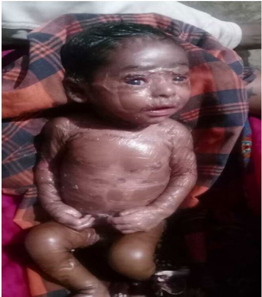
A 24-day old male baby presented to us with bilateral upper and lower lid ectropion and scaling of skin. He was born to a 28-year-old mother, gravida -2 by normal vaginal delivery at 38 weeks of gestation. There was no history of consanguineous marriage or similar complaints in any family member. Antenatal records suggested no history of birth trauma, exposure to radiation and any significant drug intake by mother during the period of gestation. As per the birth records, his birth weight was 2.3 kg and APGAR score was 10 at 10 minutes. The baby was covered with a transparent, dry, parchment- like shiny membrane resembling collodion at birth (Figure 1). Anthropometric measurements revealed cephalic circumference- 33 cm and chest diameter- 30 cm. The baby had immediately cried after birth and showed no signs of respiratory distress with a heart rate of 156 beats per minute, and respiratory rate of 45 breaths per minute. However, he had been immediately transferred to NICU in view of collodion. There, he was managed in a strictly sterile environment with adequate intravenous hydration and humidification in an incubator under the supervision of a neonatologist.
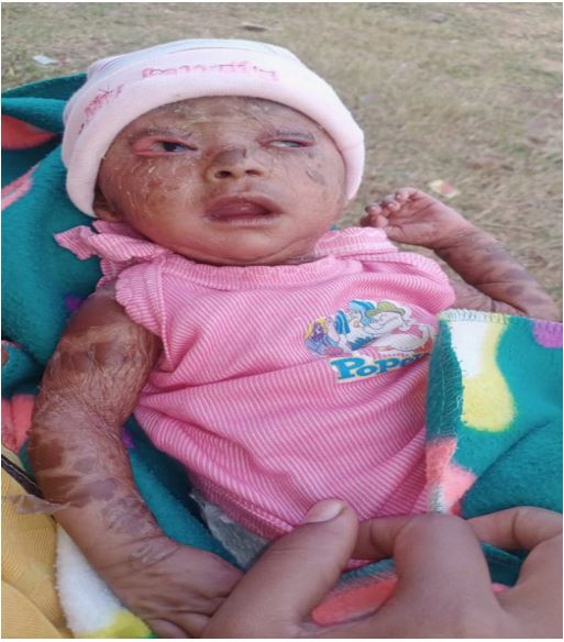
On presentation to us after 24 days of birth, there was marked bilateral cicatricial ectropion of both upper and lower eyelids with presence of bilateral eyebrows and eyelashes (Figure 2). Lid closure was incomplete and Bell’s phenomenon was good. On anterior segment examination, cornea appeared healthy without any evidence of infection. This prompted us to initiate conservative management with hourly instillation of methyl cellulose 0.5% drops, hydroxy propyl methyl cellulose 2% eye ointment every 4 hours for aggressive lubrication and 0.3% tobramycin eye drops every six hours to prevent infection. The mother was demonstrated massage of eyelid skin to be done thrice daily with same ointment and use of wet saline gauze to cover the eyes. Large brown plate like scales were seen over scalp, neck, trunk, bilateral upper and lower limbs relatively sparing face. Peeling off was evident on the entire body. The baby had been started with skin emollients for skin softening and moistening along with systemic retinoids (acitretin 10 mg dissolved in 5 ml milk) by dermatologist. Frequent follow ups were done.
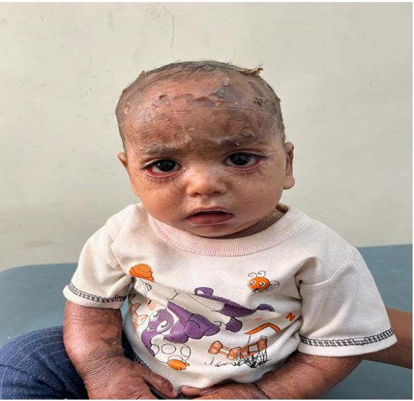
After 7 months of birth, the condition improved with remaining lower lid ectropion, while the upper lid ectropion had resolved (Figure 3). Diffuse skin peeling from multiple body sites was noted. The mother was advised to continue the same treatment and have frequent follow-ups.
Discussion
Collodion baby is an infrequent clinical presentation of numerous genetic conditions, particularly disorders of cornification [4]. Most commonly these babies have autosomal recessive inheritance owing to mutations in genes TGM 1, ALOXE3, or ALOX12B [5]. At birth, the collodion baby presents with a parchment like shiny tight skin covering the whole body. The tight skin causes mechanical compression leading to alteration of facial features and extremities. Ectropion, eclabium, pseudo contractures, absence of eyebrows, sparse hair and hypoplasia of nasal and auricular cartilage encountered in these babies are a result of the distortion [6]. The impaired skin barrier function puts these babies at a risk for various complications, including dehydration, hypothermia, infections and sepsis [7,8]. Advances in neonatal care have significantly improved the prognosis of these babies. Hence, prompt initial diagnosis and multidisciplinary involvement can optimize the outcomes.
In our case, the baby was referred for ophthalmologist opinion 24 days after birth, when ectropion became very evident. Cicatricial ectropion is the most common ocular abnormality of collodion baby. Ectropion of the lower eyelid, upper eyelid restriction, lack of Bell’s phenomenon, and eyelash retraction are assumed to cause exposure keratopathy in severe cases. In our patient no signs of exposure keratopathy were seen despite the presence of ectropion and lagopthalmos. A good Bell’s phenomenon along with upper eyelid movement could have attributed to this. Hence, conservative management was considered to deal with ectropion and lagophthalmos. Early initiation of oral acitretin, a second generation oral retinoid was also highly beneficial in our case. Acitretin induces keratinocyte differentiation in epidermal layers and accelerates shedding of hyperkeratotic skin [9,10]. It also has additional anti-inflammatory, anti-neoplastic and wound-healing effects. The optimum results obtained with conservative management, as in this case highlights the importance of early diagnosis and prompt ophthalmology care in these babies. Sight threatening complications like exposure keratopathy, corneal perforation and the need for surgical interventions could thus be averted.
Conclusion
In conclusion, a clinical diagnosis of collodion baby based on phenotype alone is sufficient instead of a definitive diagnosis. Owing to the rare occurrence of this condition, all physicians are not conversant with the treatment recommendations. However, the management is mostly supportive and requires a multidisciplinary team approach involving neonatologist, dermatologist, ophthalmologist and physiotherapist to prevent mortality and long-term complications. Affected families should also be offered genetic counselling.
Declarations
Sources of funding: Nil.
Acknowledgements: Not applicable.
Informed consent: Informed consent was taken from the patient’s father.
References
- Srivastava P, Srivastava A, Srivastava P, Betigeri AV, Verma M. Congenital Ichthyosis - Collodion Baby Case Report. J Clin Diagn Res. 2016; 10(6): SJ01-2.
- Monsudi K, Ayannil A, Teslim L. Non-surgical management of bilateral ectropion in a 5 hours old Collodion Baby:A case report. Ophthalmology Research: An International Journal. 2014; 2(5): 227–32.
- Simalti AK, Sethi H. Collodion Baby. Med J Armed Forces India. 2017; 73(2): 197-199.
- Gupta AK, Patel K, Nathwani Y, Amin N, Makwana K. Congenital bilateral ectropion in Collodion Baby: A rare case report. Delhi J Ophthalmol. 2019; 29(4): 90-2.
- Quazi S, Singh A, Khan K, Biyani U. A case report of a collodion baby: An autosomal recessive genodermatosis. Cureus. 2023; 15(4): e37418.
- Huang JJ, Huang MY, Huang TY. Lamellar ichthyosis with severe bilateral ectropion and self-healing collodion membrane. Biomarkers and Genomic Medicine. 2013; 5(3): 110–2.
- Soqia J, Mohamad L, Aloqla NA, Al Mouallem MM, EID MN. Collodion baby with ectropion in a Syrian newborn: A case report study. Annals of Medicine & Surgery. 2023; Publish Ahead of Print. doi:10.1097/ms9.0000000000000382.
- Kaur S, Kaur G, Rawat HC, Sethi A. Collodion Baby: A case report. Int J Contemp Paediatr. 2018; 5(5): 2017-19.
- Bilateral ectropion in Collodion Baby - three case reports. IP Int J Ocul Oncol Oculoplasty. 2020; 4(1): 61–3.
- Chakraborti C, Tripathi P, Bandopadhyay G, Mazumder D. Congenital bilateral ectropion in lamellar ichthyosis. Oman J Ophthalmol. 2011; 4(1): 35-6.