
Journal of Clinical Images and Medical Case Reports
ISSN 2766-7820
Clinical Image - Open Access, Volume 4
Steri-strips scaffolds: A technical tip for patients with thin skin
Ahmed Elfaki; Isabella Stevens-Harris*; Wareth Maamoun
University Hospitals of North Midlands, NHS Trust, UK.
*Corresponding Author : Isabella Stevens-Harris
University Hospitals of North Midlands, NHS Trust,
UK.
Tel: 07785395864, Fax: 07785395864;
Email: isabella.stevens-harris@nhs.net
Received : Nov 22, 2023
Accepted : Dec 21, 2023
Published : Dec 28, 2023
Archived : www.jcimcr.org
Copyright : © Stevens-Harris I (2023).
Citation: Elfaki A, Stevens-Harris I, Maamoun W. Steri-strips scaffolds: A technical tip for patients with thin skin. J Clin Images Med Case Rep. 2023; 4(12): 2762.
Description
Ageing results in changes in the collagen and elastin content within the dermis, leading to the progressive loss of integrity and elasticity. Patients with thin skin are more susceptible to significant tissue loss following minor trauma, making it more challenging to suture and prone to tearing.
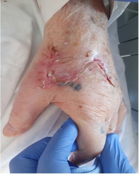
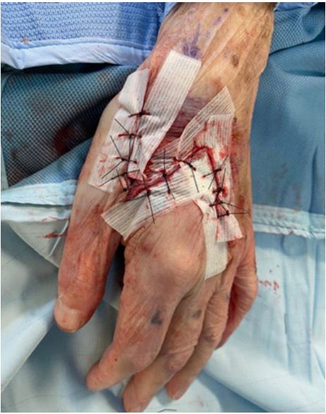
We propose the use of steri-strips in this cohort of patients to act as an additional layer to provide the skin with the strength to withstand suturing and can be utilised in both acute and elective settings. Steri-strips are often used as an alternative to suturing in patients with thin skin. Here, we have used steri-strips which are laid parallel to the edges of a wound.
This aims to provide thin skin with a scaffold and additional strength to withstand suturing without causing further damage. In our experience, this reinforced skin is less likely to tear as it is supported by the steri-strips which withstand the tension applied by the suture.
The benefit of laying the steri-strips parallel to the wound edges is two-fold. Firstly, the steri-strip can be removed easily along the length of the wound edge and therefore less likely to disrupt healing compared to it being laid in a perpendicular fashion, avoiding wound dehiscence. An additional benefit is the avoidance of unnecessary tension across the wound.
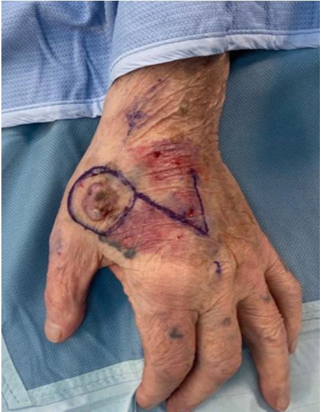
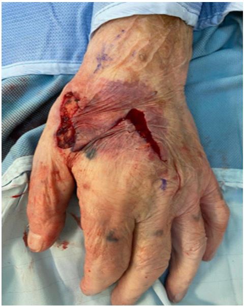
The photographs (Figures 1-4) demonstrate the case of a gentleman with a squamous cell carcinoma. Due to his thin skin, reconstruction of the defect with a skin graft would give him an additional wound with its own risk of morbidity.
The technique we describe above allowed the use of a modified Dufourmental flap, despite being thin, to reconstruct the defect following a wide local excision. This can also be used to help suture other areas of thin skin such as the pre-tibial region, where steri-strips alone can lack adequate wound apposition or where suturing alone may tear the skin. We believe that the combination of steri-strips and suturing can be a helpful technique in this cohort of patients