
Journal of Clinical Images and Medical Case Reports
ISSN 2766-7820
Clinical Image - Open Access, Volume 4
Brain abscesses: Clinical image
Catarina Alves Costa*; Ana Reinas
Internal Medicine Department, Santo António University Hospital Center, Porto, Portugal.
*Corresponding Author : Catarina Alves Costa
Internal Medicine Department, Santo António University Hospital Center, Porto, Portugal.
Email: 7catarinacosta@gmail.com
Received : Nov 23, 2023
Accepted : Dec 21, 2023
Published : Dec 28, 2023
Archived : www.jcimcr.org
Copyright : © Alves Costa C (2023).
Abstract
Brain abscesses are a rare and potentially fatal infection that can present with indolent manifestations. We present the case of brain abscesses in a 55-year-old immunocompetent man with a history of bronchiectasis, who developed significant sequalae event after appropriate antibiotic therapy and surgical drainage, highlighting the importance of early diagnosis and prompt treatment.
Keywords: Brain abscess; Pyogenic brain abscess; Central nervous system infection; Ring enhancing lesions.
Citation: Alves Costa C, Reinas A. Brain abscesses: Clinical image. J Clin Images Med Case Rep. 2023; 4(12): 2764.
Case presentation
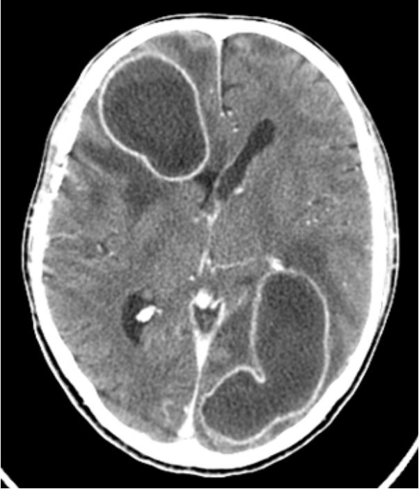
A 55-year-old man presented to the emergency department with a several-week history of fever, altered mental status and behavioral changes. He had a recent hospitalization for infected bronchiectasis. Laboratory studies revealed elevated inflammatory markers. An HIV serology was negative. A CT of the brain showed two ring enhancing lesions located in the right frontal and left temporo-occipital lobes, consistent with pyogenic brain abscesses (Figure 1). The patient underwent surgical drainage of the abscesses and completed an 8-week course of ceftriaxone, vancomycin and metronidazole. Microbiologic studies of the pus from the abscess, blood and cerebrospinal fluid were negative for bacteria, mycobacteria and fungi. After treatment, there was improvement of the mental status and behavioral changes, but the patient developed secondary epilepsy and psychosis.
Discussion
Brain abscesses are focal infections of the central nervous system [1]. These are rare infections of mainly bacterial etiology [2], more frequent in immunocompromised patients [3], that carry significant morbimortality [1,3]. Headache is the most common symptom [3]. It can be accompanied by fever and altered consciousness, but only half the patients present with focal neurological signs, depending on the abscess location [1]. Most abscesses originate in contiguous spread of a nearby infection like mastoiditis, otitis or sinusitis [3]. However, a third of cases are due to hematogenous spread from distant sites of infection as the heart or lung [3]. Appropriate antibiotic therapy in combination with surgical management are the pillars of treatment [1,3].
We report a case of brain abscesses presenting with unspecific manifestations, but with rarely seen exuberant imaging, especially in the absence of focal neurological signs. It demonstrates the high clinical suspicion required to establish this diagnosis, the need for comprehensive work-up and a prompt therapeutic approach in order to minimize sequelae. This case also highlights the often-forgotten link between lung and brain infections, even in patients with no known immunodeficiency.
References
- Ruiz-Barrera MA, Santamaria-Rodríguez AF, Zorro OF. Brain abscess: a narrative review. Neurology Perspectives. 2022; 2: 160- 167.
- Campioli CC, Almeida NE, O’Horo JC et al. Bacterial brain abscess: a outline for diagnosis and management. Am J Med. 2021; 134(10).
- Brower MC, Tunkel AR, McKhann GM, van der Beek D. Brain Abscess. NEJM. 2014; 371: 447-456.