
Journal of Clinical Images and Medical Case Reports
ISSN 2766-7820
Clinical Image - Open Access, Volume 5
A case of a large and kidney-shaped thoracic mass
Ridhi Ranchor*; Eugénia Rosendo
Medical Oncology Department, Santo António University Hospital Center, 4099-001 Porto, Portugal.
*Corresponding Author : Ridhi Ranchor
Medical Oncology Department, Santo António University Hospital Center, 4099-001 Porto, Portugal.
Tel: +351934654317;
Email: ridhiranchor@hotmail.com
Received : Nov 29, 2023
Accepted : Dec 26, 2023
Published : Jan 02, 2024
Archived : www.jcimcr.org
Copyright : © Ranchor R (2024).
Citation: Ranchor R, Rosendo E. A case of a large and kidney-shaped thoracic mass. J Clin Images Med Case Rep. 2024; 5(1): 2770.
Description
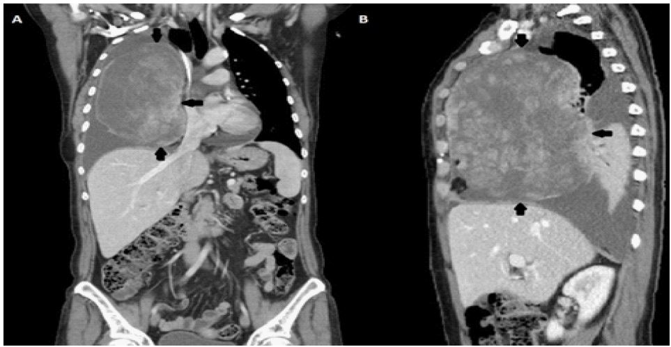
A 60-year-old woman with a high-grade myxofibrosarcoma of her left forearm, submitted to surgical excision and postoperative radiotherapy, presented to the emergency department, after 3 years, with a 2-months history of progressive asthenia and shortness of breath. On physical examination the patient had a good general condition, was acyanotic and with eupneic breathing. Cardiovascular assessment revealed a regular heartbeat with normal heart sounds, without murmurs. The peripheral oxygen saturation on room air was 94% and the vesicular murmur was absent in the entire right hemithorax. Chest computed tomography (CT) revealed a large and heterogeneous mass on the right hemithorax, measuring 13×9.4×16.4 cm, with a medium-volume pleural effusion ipsilateral (Panel 1A and B). A transthoracic biopsy was performed and the histological examination was compatible with metastasis of myxofibrosarcoma.
Following a multidisciplinary team discussion, the patient started systemic treatment with chemotherapy (doxorubicin and ifosfamide, every 3 weeks, intravenously) and was referred to the palliative care team. After 2 cycles, clinical improvement was evident, associated with a slight radiological response on CT (thoracic metastasis measuring, at this time, 12.5x9x15.5 cm). However, the patient died after these 2 cycles, due to SARS-CoV-2 infection.
Myxofibrosarcoma exhibits a high local failure rate (up to 79%), probably due to the infiltrative growth pattern. However, in some cases, distant spread can occur. The lung is the main site for sarcomatous metastasis, with multiples and well-defined nodules being one of the most common dissemination patterns. However, here we describe a case of a big, unique and bizarre sarcomatous thoracic metastasis.
Declarations
Funding: This research received no external funding.
Patient consent: Patient consent cannot be obtained because the patient cannot be traced (has died).
Conflicts of interest: The authors declare no conflict of interest.