Journal of Clinical Images and Medical Case Reports
ISSN 2766-7820
Clinical Image - Open Access, Volume 5
Re-expansion pulmonary oedema: A rare complication
*Corresponding Author : Rui Domingues
Internal Medicine, Hospital de Braga, Braga,
Portugal.
Tel: 00351916699973;
Email: ruimdf17@gmail.com
Received : Dec 15, 2023
Accepted : Jan 08, 2024
Published : Jan 15, 2024
Archived : www.jcimcr.org
Copyright : © Domingues R (2024).
Citation: Domingues R. Re-expansion pulmonary oedema: A rare complication. J Clin Images Med Case Rep. 2024; 5(1): 2796.
Description
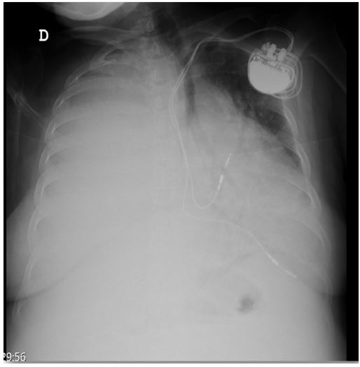
A 48-year-old female patient, with a medical history encompassing familial amyloidotic polyneuropathy and heart failure, became the focal point of a complex medical narrative. Admitted due to a symptomatic increase in right pleural effusion, accompanied by contralateral deviation of the mediastinum. In response to the clinical presentation, the medical team opted for the placement of Pleuracan®, a device designed for pleural drainage. Within the initial hour of the procedure, a notable 1400 cc of citrine yellow fluid were drained, providing a vivid insight into the urgency and extent of the pleural effusion. Surprisingly, on the same day, an additional 1500 cc were drained after the discontinuation of the procedure. The narrative took an unexpected turn as the patient developed dyspnea, necessitating oxygen therapy with a Fraction of Inspired Oxygen (FiO2) set at 60%. Seeking a more comprehensive understanding of the evolving clinical picture (Figure 1), a thoracic CT angiography was performed. The results uncovered changes suggestive of re-expansion pulmonary edema, affecting nearly the entire right lung.
In conclusion, this case offers a glimpse into the intricate interplay of preexisting medical conditions, therapeutic interventions, and the emergence of uncommon complications. Celebrating the positive outcome in this instance prompts reflection on the dynamic nature of medical care and the ongoing pursuit of knowledge to optimize patient outcomes.