
Journal of Clinical Images and Medical Case Reports
ISSN 2766-7820
Clinical Image - Open Access, Volume 5
Thymoma unveiled by right atrium cavity mass: A rare clinical image
Mehdi Belhakim1*; Jihad Aslaoui2; Hind M’chanter2; Kenza Gourram3; Evrard Niyonkuru4; Daoud Bentaleb3; Zineb Bouchbika2; Mehdi Karkouri4; Rachida Habbal1
1Cardiology Department, University Hospital Ibn Rochd, University Hassan II, Casablanca, Morocco.
2Oncology Departement, University Hospital Ibn Rochd, University Hassan II, Casablanca, Morocco.
3Radiology Department, University Hospital Ibn Rochd, University Hassan II, Casablanca, Morocco.
4Anatomopathology Department, University Hospital Ibn Rochd, University Hassan II, Casablanca, Morocco.
*Corresponding Author : Mehdi Belhakim
University Hospital Center Ibn Rochd, Hospital Street, 20360 Casablanca, Morocco.
Tel: +212-631477536 & +212520223694;
Email: mbelhakim10@gmail.com
Received : Dec 19, 2023
Accepted : Jan 11, 2024
Published : Jan 18, 2024
Archived : www.jcimcr.org
Copyright : © Belhakim M (2024).
Keywords: Right atrium mass; Thymoma; Cardiology; Oncology.
Citation: Belhakim M, Aslaoui J, M’chanter H, Gourram K, Niyonkuru E, et al. Thymoma unveiled by right atrium cavity mass: A rare clinical image. J Clin Images Med Case Rep. 2024; 5(1): 2804.
Description
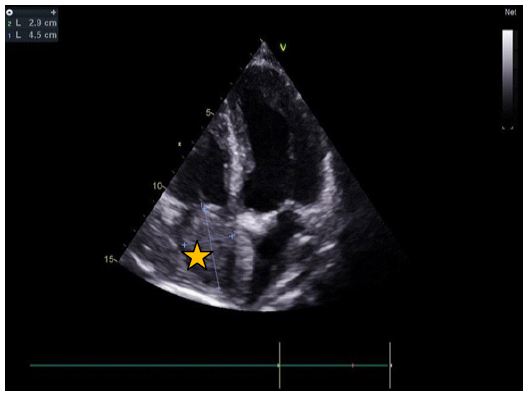
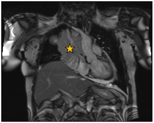
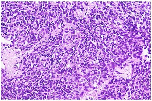
Cardiac tumors are rare, and metastatic deposits are more common than primary cardiac tumors [1,2]. We present a rare case of a 52-year-old, without significant past medical history, initially reported intermittent right shoulder pain six years ago. Recently, she developed abdominal discomfort and dyspnea, leading her to seek cardiological consultation. Echocardiography revealed a right atrium mass (Figure 1). A CT scan of the thorax disclosed a locally advanced mediastino-pulmonary mass on the left side, intimately associated with major vessels and the pericardium. Cardiac magnetic resonance imaging demonstrated a mobile anterior mediastinal mass invading the right atrium, with a preserved left ventricular ejection fraction of 69% (Figure 2). Histopathological analysis confirmed a poorly differentiated and invasive tumor, morphologically consistent with a thymoma, specifically a type B1 (Figure 3). The case was discussed at a multidisciplinary consultation meeting. The decision was to use concomitant radio chemotherapy. The patient received 60 Gray of conformal radiotherapy with intensity modulation in 30 fractions of 2 Gray, combined with concomitant chemotherapy such as cisplatin 40 mg weekly. As of the latest follow-up on June 21, 2023, the patient remained asymptomatic, with a performance status of 0. A comparative thoracic CT exhibited a 50% reduction in tumor volume. The echocardiographic control shows the disappearance of the right intra-atrial mass. This case underscores the diagnostic and therapeutic challenges posed by thymomas infiltrating the cardiac structures, emphasizing the importance of a multidisciplinary approach for effective management [3].
Declarations
Ethical approval: Written informed consent was obtained from the patient described in this article.
Funding: No funding has been received to carry out the work described in this manuscript.
Disclaimers or conflict of interest: None.
References
- Dursun M, Sarvar S, Cekrezi B, Kaba E, Bakir B, et al. Cardiac Metastasis from Invasive Thymoma Via the Superior Vena Cava: Cardiac MRI Findings. Cardiovasc Intervent Radiol. 2008; 31: 209-12.
- Kurata A, Saji H, Ikeda N, Kuroda M. Intracaval and intracardiac extension of invasive thymoma complicated by superior and inferior vena cava syndrome. Pathology International. 2013; 63: 56-62.
- De Giacomo T, Patella M, Mazzesi G, Venuta F. Successful resection of thymoma directly invading the right atrium under cardiopulmonary bypass. European Journal of Cardio-Thoracic Surgery. 2015; 48: 332-3.