
Journal of Clinical Images and Medical Case Reports
ISSN 2766-7820
Case Report - Open Access, Volume 5
Pitfalls in FDG-PET/CT: Unique brown fat activation due to a β3-adrenergic receptor agonist in a patient with treated uterine cervical cancer
Yayoi Yamamoto*; Daichi Kojima; Hiroaki Kurihara
Department of Diagnostic and Interventional Radiology, Kanagawa Cancer Center, Yokohama, Kanagawa, Japan.
*Corresponding Author : Yayoi Yamamoto
Department of Diagnostic and Interventional Radiology, Kanagawa Cancer Center, Yokohama, Kanagawa Japan.
Tel: +8145-520-2222;
Email: yamamoto.0820g@kanagawa-pho.jp
Received : Dec 26, 2023
Accepted : Jan 11, 2024
Published : Jan 18, 2024
Archived : www.jcimcr.org
Copyright : © Yamamoto Y (2024).
Abstract
Brown Adipose Tissue (BAT) activation with increased uptake on FDG-PET/CT studies is a well-known phenomenon. Activated BAT is usually seen in the supraclavicular region, but also in Para spinal and mediastinal locations, and rarely in perirenal sites. Here, we report a unique case of atypically intense, multilocular FDG uptake in activated BAT. Chart review revealed that the patient was receiving Vibegron, a β3-adrenergic receptor agonist prescribed for overactive bladder. Methods of reducing BAT uptake have been established, but there is minimal information on the pharmacologic causes of increased uptake. Factors increasing FDG uptake in BAT should be considered when interpreting FDG-PET/CT studies.
Keywords: Brown Adipose Tissue; FDG-PET/CT; β3-Adrenergic Receptor Agonist; Cancer.
Citation: Yamamoto Y, Kojima D, Kurihara H. Pitfalls in FDG-PET/CT: Unique brown fat activation due to a β3-adrenergic receptor agonist in a patient with treated uterine cervical cancer. J Clin Images Med Case Rep. 2023; 5(1): 2805.
Introduction
Brown Adipose Tissue (BAT) activation with increased uptake on FDG PET/CT studies is a well-known phenomenon. Cold exposure causes the sympathetic nervous system to release norepinephrine and induce human BAT thermogenesis through consumption of fatty acids and glucose [1,2], but little information has been reported on the pharmacologic causes of increased uptake [3-6]. Here, we report a case of remarkable FDG uptake in BAT due to a β3-adrenergic receptor (β3-AR) agonist prescribed for overactive bladder.
Case presentation
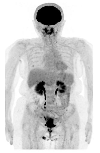
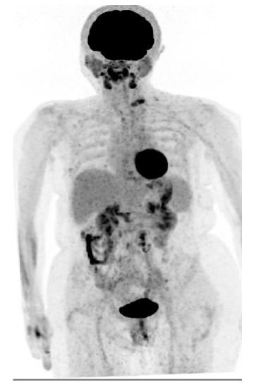
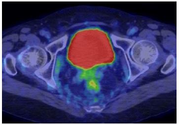
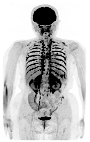
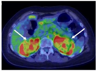
A 59-year-old female complained of lower abdominal pain. Cervical biopsy revealed uterine endocervical adenocarcinoma. CT and FDG-PET/CT showed a uterine cervical tumor and right obturator lymph node metastasis, diagnosed as T3bN1M0 (Figure 1). The patient underwent Chemotherapy with Carbon ion Radiation Therapy (CCRT). The volume of the primary tumor and lymph nodes were reduced. Six months after CCRT, tumor markers increased. FDG-PET/CT indicated metastasis to the paraaortic lymph nodes and thoracic spine (Figure 2). Chemotherapy was administered, resulting in decreased levels of tumor markers and shrinkage of lymph nodes. Ten months after CCRT, the patient suffered persistent, frequent urination and hematuria, which did not improve with antibiotics. Bladder ultrasound showed diffuse thickening of the bladder wall with no mass. Cystoscopy and cytology showed no malignancy, and the patient was considered to have an overactive bladder due to radiation cystitis. Vibegron, a β3-AR agonist, was started after no improvement was achieved with Gononsan, a type of Chinese herbal medicine. Twelve months after CCRT was complete, tumor markers were again elevated; CT failed to show any obvious recurrence and FDG-PET/CT was performed. The patient was 159 cm tall, weighed 62 kg (body mass index=24.5), and had a temperature of 29.4°C at 9:00 am on the day of FDG-PET/CT. A 180.4-MBq dose of FDG was administered at 9:59 am, at which time the blood glucose level was 103 mg/dL. Extensive distribution of FDG uptake was observed, with particularly intense uptake in the supraclavicular region. Uptake was also seen in the intercostal, paraspinal, mediastinal, retroperitoneal, and perirenal fat, which was considered to indicate BAT uptake. Uptake in the former metastatic sites, including the paraaortic lymph nodes and thoracic spine, was difficult to assess due to the BAT uptake. The intense uptake in activated BAT was thought to be due to the β3-AR agonist prescribed for overactive bladder.

Discussion
We report a case of remarkable FDG uptake in BAT due to administration of a β3-AR agonist for overactive bladder. BAT is a thermoregulatory organ that converts adipose tissue into heat. This process, called non-shivering thermogenesis, maintains body temperature under cold exposure. BAT thermogenesis is controlled by the sympathetic nervous system. Norepinephrine released from nerve terminals facilitates lipolysis in BAT by acting on β3-AR. Free fatty acids activate the specific mitochondrial thermogenic protein Uncoupling Protein 1 (UCP1), resulting in thermogenesis [3]. FDG-PET/CT has been shown to visualize activation of the BAT system in some cases. On FDG-PET/CT, BAT is most often detected in the cervical, supraclavicular, mediastinal, and paravertebral regions, and less frequently in the sub-phrenic areas. Nadia et al. reported that in patients with uptake in BAT, 93.75% had uptake in the cervical, supraclavicular, mediastinal, and thoracic paravertebral regions, and 6.25% showed uptake in the perinephric region as well. BAT is activated by cold exposure, with a higher prevalence in young, women, and lean subjects [7]. Several studies have demonstrated that tumors that secrete catecholamines, such as pheochromocytoma and paraganglioma, exhibit BAT uptake caused by elevated plasma catecholamines [8]. Vibegron, a β3-AR agonist used for overactive bladder, stimulates β3-AR in the bladder smooth muscle, causing the bladder to relax and thereby increasing urinary storage function. Activating sympathetic nerve function causes uptake in BAT, and FDG uptake in BAT due to β3-AR agonists is often stronger and more extensive than that seen in cold exposure [3-6]. The patient in the present case was middle aged and had an average body mass index, and the outside temperature was high. These conditions differed from those typically present in cases of BAT activation. FDG uptake in BAT was much stronger than usual and extended to the perirenal area, which led us to suspect drug-induced uptake. After checking the patient’s medications, we concluded that the BAT uptake was caused by the β3-AR agonist. FDG uptake in the paraaortic lymph nodes and thoracic spine, where metastases were formerly noted, was masked by the BAT uptake, making it difficult to assess. A temporary suspension of the β3-AR agonist before FDG-PET/CT is being considered in the future. In conclusion, factors increasing FDG uptake in BAT should be considered when interpreting PET/CT studies.
References
- Bartelt A, Bruns OT, Reimer R, Hohenberg H, Ittrich H, et al. Brown adipose tissue activity controls triglyceride clearance. Nat Med. 2011; 17: 200-205.
- Hondares E, Iglesias R, Giralt A, Gonzalez FJ, Giralt M, et al. Thermogenic activation induces FGF21 expression and release in brown adipose tissue. J Biol Chem. 2011; 286.
- Szentirmai É, Kapás L. The role of the brown adipose tissue in β3-adrenergic receptor activation-induced sleep, metabolic and feeding responses. Sci Rep. 2017; 7: 958.
- Brahmbhatt P, Ataei F, Parent EE, Sharma A. Atypically Intense Pharmacologically Induced Brown Fat Activation on FDG PET/CT. Clin Nucl Med. 2023; 48: 233-236.
- Okuyama C, Kikuchi R, Ikeuchi T. FDG Uptake in Brown Adipose Tissue Activated by a β3-Adrenergic Receptor Agonist Prescribed for Overactive Bladder. Clin Nucl Med. 2020; 45: 628-631.
- Cypess AM, Weiner LS, Roberts-Toler C, Elía E, Kessler S, et al. Activation of human brown adipose tissue by a β3-adrenergic receptor agonist. Cell Metab. 2015; 21: 33-38.
- Mostafa NM, Mohamadien NRM, Sayed MHD. Brown Adipose Tissue (BAT) activation at 18F-FDG PET/CT: Correlation with clinic pathological characteristics in breast cancer, Egyptian Journal of Radiology and Nuclear Medicine. 2021; 52: 62.
- Hadi M, Chen CC, Whatley M, Pacak K, Carrasquillo JA. Brown fat imaging with (18) F-6 fluorodopamine PET/CT, (18) F-FDG PET/CT, and (123) I-MIBG SPECT: A study of patients being evaluated for pheochromocytoma. J Nucl Med. 2007; 48: 1077-1083.