Journal of Clinical Images and Medical Case Reports
ISSN 2766-7820
Clinical Image - Open Access, Volume 5
A rare case of multiple myeloma of vertebral bodies with cervical canal stenosis
Harsh Rajesh Nathani; Nishigandha Deodhe*
Department of Neuro Physiotherapy, Ravi Nair Physiotherapy College, Datta Meghe Institute of Higher Education & Research (D.U.), Wardha, Maharashtra, India.
*Corresponding Author : Nishigandha Deodhe
Department of Neuro Physiotherapy, Ravi Nair Physiotherapy College, Datta Meghe Institute of Higher Education & Research (D.U.), Wardha, Maharashtra, India.
Email: nishigandha.deodhe@gmail.com
Received : Dec 27, 2023
Accepted : Jan 15, 2024
Published : Jan 22, 2024
Archived : www.jcimcr.org
Copyright : © Deodhe N (2024).
Citation: Nathani HR, Deodhe N. A rare case of multiple myeloma of vertebral bodies with cervical canal stenosis. J Clin Images Med Case Rep. 2024; 5(1): 2811.
Description
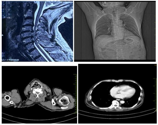
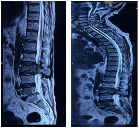
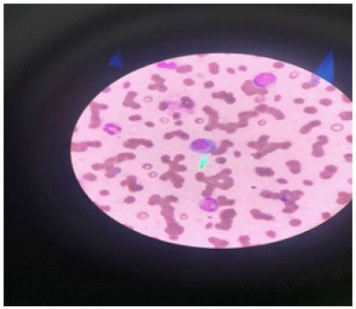
A 70-year-old male patient presented with bilateral lower limb weakness that progressively extended to the upper limbs. This weakness was accompanied by an inability to walk or stand, back pain, followed by paraparesis and paraplegia. He also experienced urine retention and no passage of stools or flatus, along with an episode of fever and weight loss. The patient reported a history of low back pain for 2 months and weight loss for 3 months. Examination revealed muscle wasting in both lower limbs, paraspinal tenderness upon palpation, and 1/5 power in all lower limb muscles. Sensation of urine passage, knee jerk, and plantar reflex were absent. Various investigations were performed- MRI of whole spine revealed- multiple metastasis of D1, D2, D6, D9, D10, D12, L1, L2, L3, L4, L5, and spinous process of L1, L2, L3 vertebrae, sacrum bilateral ileum, bilateral acetabulum also show enhancing metastatic lesion and it also suggested degenerative cervical canal stenosis at C3-C4 to C6-C7 disc level; CECT Abdomen revealed- heterogeneously enhancing soft tissue density lesion of size approx. 1.6x1 cm with erosive destruction of the body and left transverse process of the L3 vertebra. Hepatic metastasis of size 3.5x3 cm in liver parenchyma, metastasis of left adrenal body of size 1.6x1 cm and metastatic lymphadenopathy was seen. CECT Thorax revealed- Enhancing pleuroparenchymal nodules in bilateral lung parenchyma with emphysematous changes with mediastinal lymphadenopathy.
Final diagnosis: Multiple myeloma of vertebral bodies with cervical canal stenosis.
Three differential diagnosis: Monoclonal Gammopathy of Undetermined Significance (MGUS), Solitary Plasmacytoma, Waldenström Macroglobulinemia.