
Journal of Clinical Images and Medical Case Reports
ISSN 2766-7820
Review Article - Open Access, Volume 5
Implant-screw loosening: Review of the existing methods and their applications
Jacob Halevy-Politch*; Ilan Rusnak
Aerospace Engineering, Technion City, Haifa, Israel.
*Corresponding Author : Jacob Halevy-Politch
Aerospace Engineering, Technion City, Haifa, Israel.
Email: yapo@actcom.co.il
Received : Dec 18, 2023
Accepted : Jan 19, 2024
Published : Jan 26, 2024
Archived : www.jcimcr.org
Copyright : © Halevy-Politch J (2024).
Abstract
Screw Loosening (SL), called also “Implant Loosening: (IL), is a common complication after implant surgery when a screw (or screws are inserted in a bone since it was (mainly) broken, partially replaced, or cracked). Radiological approaches, both X-ray and Computed Tomography (CT) scanning are used nowadays to follow and identify this condition. Mechanical methods to study screw loosening are common in some cases (like dentistry and orthopedics); while in others (like in spinal pedicle screws) are not; Generally, these methods can be applied in a laboratory (ex-vivo) research and while extracting the screw - where screw’s condition is already known in most cases.
This paper reviews the various cases of SL as monitored by different methods that already exist and were published in the scientific literature. In the second part we propose new Ultrasonic (US) monitoring methods for in-vivo IL (or its counterpart, i.e.,” Implant Stability” (IS) - that reflects an osseointegration case of an implant).
Citation: Halevy-Politch J. Rusnak I. Implant-screw loosening: Review of the existing methods and their applications. J Clin Images Med Case Rep. 2024; 5(1): 2822.
Introduction
The skeleton is an elastic construction that enables a human (as well as an animal) to be in various static conditions (such as standing, laying, sitting) and dynamic one - walking, running, swimming, climbing, crawling, rowing a boat, pushing, lifting and much more. These positions and activities are performed in harmony with other parts of the body, like muscles, tendons, joints, and others, including nerves - enabling the ’condition’ we want to perform.
The skeleton is composed of ‘samples’-where each one of them has a specific shape and constitutes, called bones. Moreover - their constituents are different, as described in [1], according to the function they perform - like vertebras that constitute the spine; long bones; articulations (like elbow, synovial joint).
Anisotropy and heterogeneity, are characteristics belonging to the human bone [1-Ch. 2], were improved with time, and composite models were developed successfully [2,3]. At present, the most complete validation of these models was described by Cristofolini et al. [2,4], who compared synthetic bone with the human one, emphasizing the mechanical properties, such as viscoelastic behavior, main deflection, and strain distribution under axial loading, bending, and torsion for the orthopedics area strength and elasticity. In such cases, there is a requirement to improve the properties of this part (that includes the bone), or even properties of a ‘partial structure’ (i.e., that contains several parts/bones of the structure - so that it re-enabled static and dynamic conditions to be as close to the previous one.
To achieve this goal, screws are inserted in the bone -mainly for fracture fusion, but also for other reasons, as will be explained later.
It turns out that till the bone (and the skeleton) can operate again normally - as before, it requires time for healing/fusion. During this period, the screw is integrated with its surrounding bone (osseointegration) - causing better stabilization of the whole system.
It was also mentioned that the primary stability [5-7] is obtained at the surgical process, whereas the secondary one - through osseointegration phenomena, of a complex multi-time and multiscale nature (that strongly depends on the implant primary stability). Osseointegration defines the direct structural and functional connection between a living bone and the interface of a loaded carrying implant, as it was investigated by Branemark [8-10]. It was also shown [11,12] that there is good histological evidence showing that implant placement healing occurs with the bone’s intimate contact with the implant surface.
However, there are cases that the healing process does not happen, or after it was initiated, an opposite process predominates -where the bone “refuses” to integrate with the screw and it returns to its previous, failure condition (or even worse); in these cases, there is a loosening process [13-15].
In 1965 was the 1st application using the Branemark’s method for the anchorage of the fixed bridge in an edentulous jaw. Since then, millions of such and similar cases have been performed worldwide. Monitoring of the loosening process followed it.
Note: In static/rigid constructions, it is often related to pseudarthrosis (false joint) - requiring revision. Excess loading in a rigid fixation, without bone fusion, may cause implant breakage.
Methods for measuring/monitoring
Mechanical measurements- Pull-out and Torque
Measuring devices: The mechanical behavior of implants, inserted in a substrate, is obtained by measuring the pullout strength and the relative stiffness, where the insertion torque assesses only the conditions at the time of implant installation. For measuring the insertion torque (N.cm) of a screw, a Kratos torque meter (digital) is applied, while the ‘pullout tester’ (N) is applied for its pull-out force measurements. The axial traction force moves (2 mm/min) toward the long axis of the implant through a mount implant device, which is attached to a piece adapted to a load cell.
The mobility absence of the implant during a surgical procedure is defined as “primary stability”. To obtain “the primary stability”, data on insertion torque and maximum pullout force are introduced.
For testing over time, it includes a statistical model (like one-way ANOVA*) and an inequality test, as developed by Bonferroni [16]. From these tests, one obtains materials stiffnesses with the highest values.
*One-Way ANOVA (“analysis of variance”) compares the means of two or more independent groups, to determine whether there is statistical evidence that the associated population means are significantly different.
This is essential for the maintenance of the peri-implant hard and soft tissues [17-20] and, for this reason, it is a ‘determining factor’ for complete osseointegration [21-26,28-30], but it was not found as a method to determine loosening.
Non-invasive (existing) methods
Radiography (x-ray) and Computer Tomography (CT): Let us assume that an x-ray imaging (usually from two orthogonal sides) is performed on a bone. This bone contains an implanted screw. While observing the obtained radiographic image, an initial halo sign (radiolucent line around the implant >1 mm wide) is obtained. This is followed by a double halo sign on later radiographs or CT scans - defined as screw loosening [2,13,25-27].
In AP (Antero-Posterior) views (An X-ray picture, in which the beams are passing from front-to-back), a lucent ring surrounding the screw is considered a halo zone. It is confirmed in lateral view as a tracking line along part or the entire length of the screw.
A double-halo sign is used for the diagnosis of loosening in screws. Double-halo was described as a radiolucent rim surrounding the implanted screw, framed by a rim of radio-opaque dense bone (of trabeculae).
Both signs are sensitive for the diagnosis; however, the double halo sign has more specificity for the diagnosis of loose screws than the halo zone sign.
The formation of lucency is thought to be a gap, developed by the decreased fit between the screw and the bone. When the increase of lucency spreads, it leads to decreased screws fixation.
Radiolucent zones around pedicle screws have been considered to indicate loss of fixation, delayed union, or pseudoarthrosis.
Quantifying the lucency is another challenge. Sande, et al. [31] used 1 mm width to differentiate thin and wide radiolucent zones, disregarding the length of the lucency. Tokuhashi et al. [32] used 1 mm or greater circumferential lucency around a screw from two or more digitalized plain radiographic directions.
The scientific basis for choosing 1 mm as a cut-off point is not clear.
Radiolucency along the entire length of the screw is understandingly more important than isolated lucency around the tip of the screw, where a cancellous vertebral bone accounts for not more than 20% of screw strength in the lumbar vertebra.
The key factor regarding the investigation of screw loosening is the assessment of whether a screw is loosened or not, which is traditionally based on radiological approaches.
The diagnostic criteria for loosening developed by X-ray include the radiolucent area (thicker than 1 mm) around the screw [20,33-37] and the “double halo” [20,21] defined as the presence of radiolucent area and radiopaque rim at the same X-ray.
Nevertheless, the specific details regarding X-ray criteria for loosening were not described in most papers, suggesting that the subjective viewpoints of surgeons and radiologists played an important role. In general, both X-ray and CT scans lack uniform and explicit standards.
Screw loosening, which may lead to fixation failure and therefore require a surgery revision [38].
It was found that radiographic methods are very useful; however, they are ex-vivo measurements and they strongly depend on the persons performing and analyzing the images. Therefore, there may be great differences between the measured results, and experience is a critical factor here.
Periotest: Periotest is an electronic measuring device that was developed in Germany by Siemens AG in Bensheim. It performs quantitative measurements of damping characteristics of the periodontal ligament that surrounds a tooth and therefore, the established value for its mobility [39] seems unreasonable.
The handpiece of the Periotest contains an accelerated metallic slug which is directed toward the tooth under test, using an electromagnet for that purpose. The instrument measures the contact duration of the slug with the tooth, using an accelerometer, where the instrument measures ‘contact time’ versus tooth mobility. The results are displayed audibly and digitally, using a scale of (-8) for low mobility to (+50) for high mobility.
There were performed many clinical observations with Periotest, many of them are described in [44].
It was mentioned also that 6 months of Periotest measurements may indicate implant stability; it is unclear what parameters can be related to osseointegration, or vice versa to loosening.
Although the Periotest made an advantage over the existing methods at that time, including radiographic one [39], a healthy implant surrounded by a bone, will exhibit quite different stiffness characteristics and effective mass, in comparison with a tooth supported by a periodontal ligament and should appear much stiffer than shown by Periotest values.
High frequency induced mechanical vibration: Kaneko [40,41] and Kaneko, et al. [42] suggested a different, non-invasive method, for testing the integrity of the implant-tissue interface. High frequency mechanical vibrations were applied to the tested implant. This method required penetration of puncture needles through the mucosa to the implant, for transmitting the signals to it. However, it was found that the sensitivity was low to interfacial changes.
It was also stated [40] that their mechanical vibrations may provide information on the mechanical bone interface; however, the pulsed oscillations are sensitive to the direction of loading as well as to its position, which reduced in most cases their amplitude.
Resonance Frequency Analysis (RFA): Resonance Frequency Analysis (RFA) is also a non-invasive testing method that performs quantitative stability measurements of the implant-tissue interface. This method can operate in-vitro, as well as in-vivo.
The transducer in RFA is attached to the implant directly, or using a transmucosal abutment screw [43-46]. The transducer used here, behaves like a cantilever beam that vibrates freely and is supported at its end - where it is attached to the implant. For a fixed length of the transducer, the resonance frequency of the whole system depends on the length of the abutment and the bone level surrounding the implant fixture.
The resonance frequency is given [47] by:
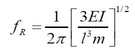
where: fR = resonance frequency [Hz], l = effective length of the beam [m]
m = mass of the beam [Kg], E = Young’s modulus [GNm-2], and I = moment of inertia [Kg m2].
From the above equation, it follows that fR varies with the implant’s stiffness in the surrounding tissue.
Generally, it is considered that there is a union between the surface of an implant surrounding the bone [43]. The term ‘osseointegration’ was defined by Branemark, et al. [8,9], where it defines a direct structural and functional connection between an ordered living bone tissue and the surface of a loaded carrying implant. Sennerby, et al. [11,12] showed that there is good histological evidence for that assumption, following implant placements, for healing of bone in contact with the implant surface.
The implant may fail due to several reasons, including trauma, infection, placement in compromised tissue, or due to overloading.
Failure is expressed usually as: (a) increasing, progressive mobility of the implant; (b) decrease in height of the surrounding marginal bone; and (c) fracture of one or more implant components.
RFA can analyze in-vivo the bone formation around an inserted implant, by using a small transducer that is attached to the fixture’s implant. The resonance frequency is changing and represents changes in the implant-tissue interface and the bone surrounding the implant fixture.
As described in many publications, the RFA method was found suitable for monitoring osseointegration. However, when considering a loosening process, the peaks of resonance become flat -as described in [5]. Thus, even if it was followed by a numerical detailed analysis and simulation- it was not possible to determine the conditions that follow the loosening process.
The resonance frequency fR [Hz] in RFA is obtained by using a small transducer, attached to the implant under consideration and headed at different heights of an Al block. A high correlation was found between fR and the height of the exposed implant fixture. It was also observed that during the healing process, there was a change in stiffness, expressed in a significant increase of fR.
Rittel D. et al. [5] analyzed by simulation the RFA method and indicated that it operates properly when there exists osseointegration, where are obtained sharp resonance peaks. However, when loosening starts to appear, the sharp peaks become flat curves -where there is no possibility to define the loosening status. Accordingly, RFA operates properly only for healthy conditions -where osseointegration is part of the healing process.
Applications
Dental
It is interesting to note that the dental methods for testing implant stability and therefore osseointegration, were the pioneering ones. They are popular among dentists performing implantations. Naturally, in the beginning, the mechanical methods (pull-out and torque) were used mainly; however, as the Periotest arrived on the market, it started to be used in more cases. It was used, as in the mechanical method, together with the ISQ (Implant Stability Quotation) [50] method [12] which defines and indicated the stability level (and therefore the osseointegration on dental implants). The ISQ [50] scale range was defined from 1 to 100, where higher values indicate better stability. For dental implants, the acceptable stability ranges between 55 and 85 of these units. Despite its success, the evaluation of a loosening process was almost impossible.
This is even though Periotest provided for the first time the advantage of in-vivo measurements (in comparison to mechanical ones).
As the radiographic methods were developed more, they started to be applied for implantations: (a) before the Implantation Surgery (IS) -for its detailed planning; (b) During the surgery (intra-operatively) – for verifying that the planned and obtained implant conditions within the inserted bone, are similar - if not the same. For such a purpose, there may be taken two perpendicular images, or from two angles which are as close as possible to 90o; and (c) During the healing process. In this case, there was the first time that thin halos were observed in both radiographic projected images. These halos were just in the thin soft material, located between the implant (screw) and the trabecular bone (which is described and presented in [5] and many others). The advantage in this method that it is possible to follow after the mentioned halos, by performing such radiograph images at defined times and measuring the halos. If they become thinner with time -there is an osseointegration; however, if they become wider -there is a loosening.
There are some disadvantages related to this method: (a) The patient receives a relatively high dose of x-ray radiation over time; (b) the interpretation of the results is dependable on the person that provides the measurements on the radiographs - his skills and experience; (c) although it is a non-invasive method, and can be used in some cases in-vivo, it is not an intraoperative one, i.e. the surgery has to be stopped to perform the radiograph.
A different method was developed by Meredith N. et al. [43-46], in which quantitative stability of the implant-tissue interface was obtained, using the RFA measuring principle. To perform this kind of measurement, it is required to attach to the implant a metallic bar and according to its vibrations define the resonance frequencies – thus the implant stability and the status of osseointegration. It was found that the accuracy of this method is a function of the width of the peak in the resonance; i.e., sharp for high stability. However, as the stability is lower, the peak of the resonance frequency is broader, thus defining it becomes almost impossible in most cases. This problem was demonstrated already in [5].
This method can be applied in-vivo and does not require special training for its application and most importantly - the results are reproducible and independent of the person performing the measurement [48].
There were developed also other RFA methods, as described by Valderma [49], and the calculation of the ISQ for a laser scanning vibrometer system - by Debtiyne S. et al. [50].
Craniofacial and bone-anchored hearing aids (BAHA) and particular prosthesis: In craniofacial implant surgeries, there is a concern, among others, with the following subjects: temporal bone, periorbital bone, and nose, as well as Bone-Anchored Hearing Aids (BAHA) and articular prostheses.
In the past, mechanical methods were applied, followed by Periostat measurements; nowadays RFA is mostly applied in these cases, as it provides the in-vivo measured information of the implant’s stability. As mentioned, implant stability is correlated with the osseointegration stage of the bone surrounding it.
When there is an interest in providing the sensitivity and monitoring changes in implant stability, a follow-up is required, including stability measurements of the implant at every measuring condition. It was suggested [51,52] that these measuring observations should be performed at defined periods - to minimize the statistical error.
In clinical-implant-surgery-studies, where there is a follow-up for many years (up to15 years) of hundreds of patients, are applied definitions that help to arrive at conclusions, on the same statistical basis and the statistical error, while assessing stability during each stage of analysis. This is even if there was performed in a different follow-up (whereas in many cases Student’s t-test is used for the statistical analysis).
Accordingly, a ‘worst case scenario’ assumes that all patients lost to follow-up, deceased patients, and those who left the study were failures are also included.
All these terms are according to [53,54] and are deemed as follows:
(1) Failure. Implants lost due to loss of integration or due to direct trauma towards the implant.
(2) Implants in dead patients. The number of implants in patients who died during a specific time after the insertion of the implant.
(3) Number of implants lost to follow-up. Implants in patients who have not taken part in the out-patient follow-up schedule.
(4) Number of implants who have left the study. Patients who removed their implants for some reason during the study.
(5) Number of implants withdrawn. The sum of implants lost to follow-up, plus implants in diseased patients, plus implants in patients who have left the study.
(6) Annual failure rate. The number of failures divided by the number of inserted implants minus half of the implants withdrawn.
(7) Annual success rate. 100 minus annual failure rates.
(8) Survival rate. The cumulative annual success rate.
(9) ‘Worst case’ success rate. Number of implants minus failures minus implants withdrawn divided by the number of implants.
(10) ‘Worst case’ cumulative success rate. The cumulative ‘worst case’ success rate.
Final remarks: (a) It was found that early and late implant failures occurred often in patients that received irradiation in the region where implants were placed [69]. (b) It was also found that implants placed in maxillary and periorbital bone for many years (can achieve even 15 years), a lower success was obtained than in the temporal bone [70-72], due to different bone quality.
In conclusion: by longer healing of the implanted soft bone, better stability was achieved. Similar results were obtained, for dense bones, with short healing.
Orthopedics
Implants are frequently used in orthopedics when a bone, or a part of it, has to be replaced, or due to a fracture (or a crack) of it. In most cases, the implants in orthopedics are of larger dimensions than those used in other parts of the body. Therefore, it is important for the osseointegration process to control their stability during the insertion, as well as with time (which may turn, in some cases, to loosening).
At the beginning, the mechanical tests [29,30,54] were applied (pull-out and insertion torque). These are invasive methods, dependent on the person that performs the measurement and requires also an ISQ [50] interpretation. After Ostell instrument [55] was introduced and followed by the RFA method (developed by Meredith N. et al. [44-46]), they were mainly applied in various implant surgery applications and follow-ups. In parallel, the radiographic and CT non-invasive methods continued to provide the required information, in spite their drawbacks, as mentioned earlier.
Following the Meredith N. investigation, Mikami K. et al. [55,56] developed a laser pulsed system (several Joules per pulse, based on an Nd-YAG laser), providing short pulses of 50 nsec with a PRF=50 Hz. The detection of the resonance frequency was performed by a ‘laser Doppler analyzer’. The measuring results were similar to those obtained with other non-invasive existing methods. These short and high-power laser pulses induced a ‘radiation pressure’ [58] on the screw’s elastic vibrations that enabled amplitude measurements, especially at its resonance frequencies.
Pelvis and its parts
The methods described above are used these days; however new directions are shown more frequently, due to the size and construction of the various parts in the pelvis.
The RFA remained the major analytical tool, but more applications appeared, showing the high-power pulsed laser as the preferable source of impact excitation. As mentioned, this approach was initiated by Mikami K. et al. [56,57] and further developed by him and his collaborators [59,60]. Kikuchi S. et al. [61] developed in 2019 a laser-RFA method for acetabular cup stability during surgery. It is interesting to note, that for the same purpose, but less efficiently, Henysh P. et al. [62] applied the conventional RFA.
HIP
The hip is categorized as another large bone. When there is a necessity to insert an implant, the conventional methods to follow the healing process were not adequate, and people were looking for better methods.
For example, Georgio AP, et al. 2001 [63] followed by Qi G. et al. 2003 [64] applied acoustical vibrations in the case of ‘total hip replacement’. They were interested to learn more about the loosening process that may occur in such surgeries.
Another approach to obtain implant stability of a cementless hip prosthesis was performed by producing a short mechanical impact followed by its frequency response function [65], where it was also been able to apply intra-operatively and measure the ‘primary stability’ [66].
A different approach was described in [67], for monitoring loosening in a hip prosthesis, by measuring in parallel: (1) the ‘acoustic emission’ from it and (2) a pulsed signal that was transmitted from an oscillating device, inserted in the hip. (This device was turned to operate by an external trigger).
Spine/vertebra
Mechanical methods (pull-out and torque) were used in this case as well [26-28] -similar to other methods mentioned above.
In radiologic and CT measurements, it was found that when there exists around a screw on plain radiographs from 2 or more directions a circumferential lucency of 1 mm or greater [31,32], the patient was judged as ‘clear-zone positive’.
The course of ‘clear-zone positivity’ was analyzed and the relationships between it and the following items were investigated accordingly:
(1) number of fused levels; (2) bone union; (3) posterior-lateral lumbar fusion versus posterior lumbar interbody fusion; (4) clinical results; (5) bone mineral density; and (6) screw types.
It was found that clear zones persisting for 2 years or longer after surgery are a great risk of pseudarthrosis. Therefore, careful observation of clear zones around pedicle screws is of great significance as an evaluation of bone union.
Although, the presence of clear zones does not necessarily indicate the presence of pseudarthrosis; as it was found that clear zones disappeared with time in many patients, showing that the extension or disappearance of clear zones after their generation is more important than clear-zone presence.
However, as clear zones were observed in more than 90% of patients with pseudarthrosis, clear zones persisting for 2 years or longer after surgery, are highly predictive signs of pseudarthrosis.
Studies reporting on RFA applications, similar to those in dental implantology and auditory Osseo integrated implants were also identified for spine/vertebra cases and similarly, methods of monitoring.
Table 1: The course of endoscopic treatments.
| Measuring Methods and their Applicationsfor Loosening & Osseo integration. | |||||||||
|---|---|---|---|---|---|---|---|---|---|
| Measuring | Mechanical | Periotest | Radiography | RFA | Meredith N. | Ostel | Laser | Acoustical | Mechanical |
| Method | (Pull-out & Torque) | *Pre Surgery *Intraoperative | (Resonance | Method | Pulsed | Vibrations | Impact+ | ||
| *On Healing * Non-invasive | Frequency Analysis) | System | Frequency | ||||||
| *Accumulative dose of radiation | Response | ||||||||
| Dental | yes | yes | yes | yes | yes | ||||
| Ex-vivo | In-vivo | Follow-ip while healing | * Implant Stability | ||||||
| Requires ISQ (Implant Stability Quotation) | *In-vivo status of osseointegration | ||||||||
| Cranial, BAHA & | yes | yes | yes | yes | yes | ||||
| Artificial Prosthesis | * In-vivo implant | ||||||||
| stability | |||||||||
| Orthopedic | yes | yes (Nowadays) | yes | yes | yes | yes | yes | ||
| * Pelvis and its parts | yes | yes | yes | yes | yes | yes | yes | ||
| * Hip | yes | yes | yes | yes | yes | yes | yes | yes | |
| Spine/Vertebra | yes | yes | yes | ||||||
Conclusion
This review describes the evolution of loosening measurement and monitoring and the way that they were applied to the different parts of the human skeleton. As can be seen above, there are nowadays, in some cases, more than one method to monitor loosening; however, each one has its drawback that prevents obtaining reliable results ex-vivo, or require a high dosage of accumulated x-ray (for following the loosening process over a long period, of years). Interesting to note that there is no ultrasonic method for this purpose. Such methods we have investigated and will be described in our next paper.
Acknowledgment: The authors are most thankful to Professor Dan Adam, Dept. of Biomedical Eng., Technion I.I.T., for his comments while preparing this review.
References
- JD Currey, BONES, md Ed. Princeton Univ. Press. 2002; 10: 0-691-1280-9
- L. Cristofolini, M. Visconti, Mechanical validation of whole bone composite tibia models, Journal of Biomechanics. 2000; 279-288.
- MA Carvalho, CM Queiroz, CCL Molena, CP Rezende, A Rapoport. Clinical study of the relationship between the implant insertion torque and the osseointegration, Revista Brasileira de Cirurgia de Cabeça e Pescoço. 2008; 202-205.
- L Cristfolini, M Visconti, A Cappello, A Toni. Mechanical validation of whole bone composite femur models, Journal of Biomechanics. 1996; 525-535.
- D Rittel, A Dorogoy, G Haïat, K Shemtov-Yona. Resonant frequency analysis of dental implants Medical Eng. And Physics. 2019; 65-77.
- NF Olisovicz, et al. Analysis of Primary Stability of Dental Implants Inserted in Different Substances, using the Pullout Test and Insertion Torque, Intl’ J. of Dentistry. 2013.
- Yamaguchi K, Konishi H, Hara S. Histological study of titanium pediclescrew/tissue interface. J Jpn Soc Spine 1996; 7: 131.
- Bra°nemark PL. Prostheses Tissue Integrated, Amazon 01, 03, 1985. Note: R. Bra°nemark research has been made in 1952, during his study on healing reaction of Titanium screw in a rabbit bone. 1985.
- Bra°nemark PL, Hansson BQ, Adell R, Breine U, Lindstrom J, et al. Osseointegrated implants in the treatment of the edentulous jaw. Scandinavian Journal of Plastic and Reconstructive Surgery. 1977; 111: 1-132.
- Del Valle V, Faukner MG, Wolfaardt J, Rangert B, Tan H. Mechanical evaluation of craniofacial osseointegration retention systems. Department of Mechanical Engineering, University of Alberta, Edmonton, CanadaDepartment Report Number. 1993; 88: 1-28.
- Sennerby L, Thomsen P, Ericson LE. Early bone tissue responses to titanium implants inserted in rabbit cortical bone (1). Light microscopic observations. Joumal of Materials Science Materials in Medicine. 1993; 4: 240-250.
- Sennerby L, Meredith N. Stability measurements using resonance frequency analysis: Biological and biomechanical aspects and clinical implications. Implant Periodontol 2000. 2008; 47: 55-66.
- Ko CC, Tsai HW, Huang WC, Wu JC, Chen YC, et al. Screw loosening in the Dynesys stabilization system: Radiographic evidence and effect on outcomes. Neurosurg Focus. 2010; 28:10.
- Galbusera F, Volkheimer D, Reitmaier S, Berger-Roscher N, Kienle A, et al. Pedicle screw loosening: A clinically relevant complication?. Eur Spine J. 2015; 24: 1005-16.
- Wu JC, Huang WC, Tsai HW, Ko CC, Wu CL, et al. Pedicle screw loosening in dynamic stabilization: Incidence, risk, and outcome in 126 patients. Neurosurg Focus. 2011; 31: 9.
- Hayer. What Is the Bonferroni Test (Correction) and How Is It Used?. Fundamental Analysis/Tools. 2021.
- Dakhil-Jerew F, Jadeja H, Cohen A, Shepperd JA: Inter-observer reliability of detecting Dynesys pedicle screw using plain X-rays: A study on 50 post- operative patients. Eur Spine J. 2009; 18: 1486-1493.
- Bayarchimeg D, Namgoong H, Kim BK, Kim MD, Kim S, et al. Evaluation of the correlation between insertion torque and primary stability of dental implants using a block bone test. J Periodontal Implant Sci. 2013; 43: 30-6.
- Dorogoy A, Rittel D, Shemtov-Yona K, Korabi R. Modeling dental implant insertion. J Mech Behav Biomed Mater. 2017; 68: 42-50.
- S Kahraman, BT BAL, NV Asar, I Turkyilmaz, TF Tözüm. “Clinical study on the insertion torque and wireless resonance frequency analysis in the assessment of torque capacity and stability of self-tapping dental implants,” Journal of Oral Rehabilitation. 2009; 755-761.
- S Inceoglu, L Ferrara, RF McLain. Pedicle screw fixation strength: Pullout versus insertional torque, Spine Journal. 2004; 513-518.
- VC Leite, AC Shimano, GAP Gonçalves, F Kandziora, HLA Derno. The influence of insertion torque on pedicular screws pullout resistance. Acta Ortopedica Brasileira. 2008; 214-216.
- RC Rosa, P Silva, AC Shimano, JB Volpon, HLA DeFno, et al. Biomechanical analysis of the variables related to the pullout strength of screws in the vertebral fixation system. Revista Brasileira de Ortopedia. 2008; 293-239.
- KK Salmória, OM Tanaka, O Guariza-Filho, ES Camargo, LT de Souza, et al. Insertional torque and axial pullout strength of mini-implants in mandibles of dogs. American Journal of Orthodontics and Dentofacial Orthopedics. 2008.
- WC Tsai, PQ Chen, TW Lu, SS Wu, KS Shih, et al. Comparison and prediction of pullout strength of conical and cylindrical pedicle screws within a synthetic bone. BMC Musculoskeletal Disorders. 2009.
- Yamaguchi K, Konishi H, Hara S. Histological study of titanium pedicle screw/tissue interface. J Jpn Soc Spine 1996; 7: 131.
- Yamaguchi K, Konishi H, Hara S, et al. Histological study of a titanium pedicle screw/tissue interface 2; Effect of anterior fixation on pedicle screw loosening. J Jpn Soc Spine 1997; 8: 262.
- A Camarioli, PA Simches, AC Shimano, HLA DeRno. Insertion torque and pullout strength of vertebral screws with cylindrical and conic core. Revista Brasileira de Ortopedia. 2008; 452-459.
- L Cristofolini, M Viceconti, A Cappello, A Toni. Mechanical validation of whole bone composite femur models. Journal of Biomechanics. 1996; 525-535.
- L Cristofolini, M Viceconti. Mechanical validation of whole bone composite tibia models. Journal of Biomechanics. 2000. 279-288.
- Sandén B, Olerud C, Petrén-Mallmin M, Johansson C, Larsson S. The significance of radiolucent zones surrounding pedicle screws. Definition of screw loosening in spinal instrumentation. J Bone Joint Surg Br. 2004; 86: 457-461.
- Tokuhashi Y, Matsuzaki H, Oda H, Uei H. Clinical course and significance of the clear zone around the pedicle screws in the lumbar degenerative disease. Spine. 2008; 33: 903-908.
- A Rutar, NP Lang, D Buser, W Bürgin, A. Mombelli. Retrospective assessment of clinical and microbiological factors affecting peri-implant tissue conditions. Clinical Oral Implants Research. 2001; 189-195.
- JEIG Brouwers, F Lobbezoo, CM Visscher, D Wismeijer, M Naeije. Reliability and validity of the instrumental assessment of implant stability in dry human mandibles. Journal of Oral Rehabilitation. 2009; 279-283.
- P Trisi, W Rao. Bone classification: Clinical-Histomorphometric comparison. Clinical Oral Implants Research. 1999.
- L Chong, A Khocht, JB Suzuki, J Gaughan, Effect of implant design on initial stability of tapered implants. Journal of Oral Implantology. 2009; 130-135.
- MC Cehreli, D Karasoy, K Akca, SE Eckert. Metaanalysis of methods used to assess implant stability. International Journal of Oral & Maxillofacial Implants. 2009; 1015-1032.
- JS Hermann, D Buser, RK Schenk, JD Schooltheld, DL Cochran. Biologic Width around one- and two-piece titanium implants-a histometric evaluation of unloaded non-submerged and submerged implants in the canine mandible. Clinical Oral Implants Research. 2001; 559-571.
- Schulte W, Lucas D, Mtihlbradt L, Scholz F, Bretschi J, et al. Periotestein neues Verfahren un Gerat zur Messung der Function des Parodontiums. Zah niirtzl Mitt. 1983; 73: 1229.
- Kaneko T, Nagai Y, Ogino M, Futami T, lchimura T. Acoustoelectric technique for assessing the mechanical state of the dental implant-bone interface. Journal of Biomedical Materials Research. 1986; 20: 169-176.
- Kaneko T. Assessment of the interfacial rigidity of bone implants from vibrational signals. Journal of Material Science. 1987; 22: 3495-3502.
- Kaneko T. Pulsed oscillation technique for assessing the mechanical state of the dental implant-bone interface. Biomaterials. 1991; 12: 555-560.
- Meredith N, Cawley P, AJleyne D. The application of modal vibration analysis to study bone healing in-vivo. Jounal of Dental Research. 1994; 73: 793.
- Meredith N, et al. Quantitative determination of the stability of the implant-tissue interface using resonance frequency analysis. Clin. Oral Implant Res. 1996; 7: 261-267.
- Meredith N, et al. Resonance frequency measurements of implant stability in- vivo. A cross-sectional and longitudinal study of resonance frequency measurements on implants in the edentulous and partially dentate maxilla. Clin. Oral Impl. Res. 1997; 8: 226-233.
- Meredith N, et al. The application resonance frequency measurements to study the stability of Titanium implants during healing in the rabbit tibia. Clin. Oral Impl. Res. 1997: 8: 234-243.
- Timoshenko S, Goodyear JN. Theory of Elasticity. 2nd Ed. McGra-Hill.1984.
- Huwiler MA, et al. Resonance frequency analysis in relation to jaw bone characteristics and during early healing of implant installation. Clin Oral Implant Res. 2007; 18: 275-280.
- Valderma P, et al. Estimation of two different resonance frequency devices to detect implant stability: clinical trials. J. Periodont. 2007; 78: 262-272.
- Debtiyne S, et al. ISQ (Implant Stability Quotation) calculation evaluation of in-vitro laser scanning vibrometry- captured resonance frequency. Intl’ J. Implant Dent. 2017; 3-44.
- Heo SH, et al. Stability measurements of craniofacial implants by means of resonance frequency analysis: A clinical pilot study. The Journal of Laryngology and Orology. 1998; 112: 537-542.
- Tjellstrom A, et al. One stage procedure to establish osseointegration: A zero to five years follow-up report. The J. Laryngology and Otology. 1995; 109: 593-598.
- Armitage P, Berry G. Statistical Methods in Medical Research, 2nd Edition, Ch.14, Blackwell Scientific, Oxford. 1987; 421-439.
- Murray DW, Carr AJ, Bulstrode C. Survival analysis of joint re placements. Journal of Bone and Joint Surgery. 1993; 75: 697-704.
- Ostell. (a) User manual and (b) mentor user manual, Gothenburg, Integration Diagnostics AB.
- Mikami K, et al. Flash-Lamp-Pumped 4J, 50 Hz, Nd-YAG nanosecond laser system for mobile and transportable equipment. 2017.
- Mikami K, et al. Stability diagnosis of orthopedic implants based on resonance frequency analysis with fiber transmission of nanosecond laser pulse and acceleration sensor. Conf. Paper 2/2020: Optical Fiber and Sensors for Medical Diagnostics and Treatment Applications. 2020.
- Mahrle A, Dayer E. Theoretical evaluation of radiation pressure magnitudes and effects in laser materials processing. Phus. 2019.
- Nakashima D, Mikami K, et al. Laser resonance frequency analysis of pedicle screw stability: A cadaver model bone study. J. of Orthopedic Res. 2021; 39.
- Mikami K, et al. Sweep-pulse excitation method of enhancing photoacoustic elastic waves at different laser irradiation parameters. Sensors. 2022; 22.
- Kikuchi S, et al. Laser resonance frequency analysis measurement approach to evaluate acetabular cup stability during surgery, Sensors. 2019; 19: 48-71.
- Henysh P, et al. Evaluation of acetabular cup initial fixation by using resonance frequency principle. Proc. Inst. Mech. Eng, Part H: J. of Engineering in Medicine, Feb. 2015.
- Georgio AP, et al. Accurate diagnosis of hip prosthesis loosening using vibrational technique, Clin Biomech. 2001; 16: 315-323.
- Qi G, et al.How much can a vibrational diagnostic tool reveal in total hip arthroplasty loosening?. Clin Biomech. 2003; 18: 444-458.
- Varini E, et al. Assessment of implant stability of cementless hip prosthesis through frequency response function of stem bone system. Sens Actuator: A Phys z010B 165: 526-532.
- Cristofolini E, et al. Device to measure intra-operatively primary stability of cementless hip stems. Med. Eng. Phys. 2006; 28: 475-482.
- Ruther C, et al. Investigation of acoustic mechanical method to detect implant loosening. Med. Eng. Phys. 2013; 35: 1669-1675.
- Zindrick MR, Wiltse LL, Widell EH, et al. A biomechanical study of intra- peduncular screw fixation in the lumbosacral spine. Clin Orthop 1986; 203: 9-112.
- Granström G, Bergström K, Tjellströom A, Brånemark PI. A detailed analysis of titanium implants lost in irradiated tissues. International Journal of Oral and Maxillofacial Implants. 1994; 9: 653-662.
- Granströom G, Tjellström A, Brånemark PI, Fornander J. Bone-anchored reconstruction of the irradiated head and neck cancer patient. Otolaryngology-Head and Neck Surgery. 1993; 108: 334-343.
- Roumanas E, Nishimura R, Beumer J, Moy P, Weinlander M, et al. Craniofacial defects and Osseo integrated implants: Six-year follow-up report on the success rates of craniofacial implants at UCLA. International Journal of Oral and Maxillofacial Implants. 1994; 9: 579-585.
- Faber HT, et al. Bone anchored hearing implant loading at 3 weeks Stability and tolerability after 6 months. Otology and Neurology. 2012; 34: 104-110.