
Journal of Clinical Images and Medical Case Reports
ISSN 2766-7820
Clinical Image - Open Access, Volume 5
Recurrent rhinosporidiosis: A clinical picture
Prasanth L*; Jyoti Mishra; Niharika Mishra; Roohie Singh
Command Hospital (Eastern Command), Kolkata, West Bengal, India.
*Corresponding Author : Prasanth L
Command Hospital (Eastern Command), Kolkata, West Bengal, India.
Email: ragavprash28@gmail.com
Received : Jan 17, 2024
Accepted : Feb 05, 2024
Published : Feb 12, 2024
Archived : www.jcimcr.org
Copyright : © Prasanth L (2024).
Abstract
Citation: Prasanth L, Mishra J, Mishra N, Singh R. Recurrent rhinosporidiosis: A clinical picture. J Clin Images Med Case Rep. 2024; 5(2): 2854.
Description
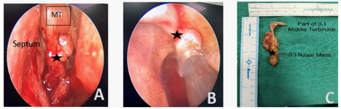
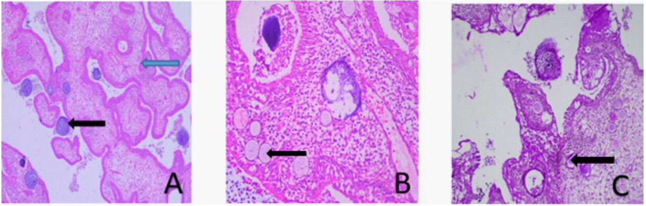
28 year, male, driver by profession and resident of Kerala presented with complaints of recurrent Left nasal obstruction and intermittent epistaxis from left nostril of 6 months. He had underwent four excisional surgeries since last 3 years for same complaints. Diagnostic nasal endoscopy revealed a strawberry like mass which bled on probing. It was filling left nasal cavity likely arising from middle turbinate (Figure 1A). CECT of paranasal sinuses revealed soft tissue attenuating content in left nasal cavity. The patient underwent Coblation assisted endoscopic excision of (L) nasal mass (Figure 1B). The mass was excised in toto with anterior part of left middle turbinate to which it was attached (Figure 1C). Histopathological examination was consistent with Rhinosporidiosis (Figure 2A-2C). Postoperatively the patient was started on Tablet Dapsone 100 mg once daily for 6 months.
Discussion
Rhinosporidiosis is a chronic granulomatous infectious disease which predominately affects the mucosal membranes of nose and nasopharynx, caused by the parasite Rhinosporidium seeberi. It usually occurs as a result of direct traumatic inoculation with the organism which grows in stagnant water. The characteristic lesions in the nasal passage are polypoidal, granular, red in colour due to pronounced vascularity, with a surface containing yellowish pin head sized spots which represents underlying mature sporangia [1].
The definitive diagnosis of Rhinosporidiosis is by histopathology with the identification of the pathogen in its diverse stages. Radiological imaging particularly CT and MRI are helpful in outlining the extent of the disease as well as excluding other pathology in patients presenting with nasopharyngeal masses. Differential diagnosis include antrochoanal polyp with squamous metaplasia, inverted papilloma, extra nasopharyngeal angiofibroma, etc. [2].
Recurrence is known to occur in 10% of cases after excision [3]. Here, we emphasize the importance of coblation assisted excision and medical management using Dapsone to reduce risk of recurrence.
Declarations
Conflicts of interest: Nil.
Informed consent: Informed consent was taken from the patient for this publication.
Funding: No funding was received to assist with the preparation of this manuscript. The authors have no relevant financial or non-financial interests to disclose.
References
- Tong TK, Ismail I, Yunus Mohammad NM, Yusoff SM, Sahri AM. Recurrent rhinosporidiosis: A case report from Malaysia and review of literature. Proceedings of Singapore Healthcare. 2023; 32: 20101058231160606.
- Venkatachalam VP, Anand N, Bhooshan O. Rhinosporidiosis: Its varied presentations. Indian Journal of Otolaryngology and Head & Neck Surgery. 2007; 59: 142-4.
- Majumdar AB, Biswas D, Paul SS, Ray S, Kumar G. Rhinosporidiosis: a clinicopathological study from a Rural Tertiary Health Care Centre, Bihar, India.