
Journal of Clinical Images and Medical Case Reports
ISSN 2766-7820
Case Report - Open Access, Volume 5
Treatment of horizontal middle root fracture with TotalFill bioceramic material: Case report
*Corresponding Author : Mohammed Abdullah Alzubaidi
Assistant Professor in Pediatric Dentistry, Department of Preventive Dentistry, Faculty of Dentistry,
Taif University, P.O. Box 11099, Taif 21944, Saudi
Arabia.
Email: m.alzubaidi@tu.edu.sa
ORCID ID: 0009-0009-4222-4719
Received : Jan 22, 2024
Accepted : Feb 09, 2024
Published : Feb 16, 2024
Archived : www.jcimcr.org
Copyright : © Alzubaidi MA (2024).
Abstract
Root fractures represent 0.5 to 7% of the overall occurrences of traumatic dental injuries that affect the permanent dentition’s cementum, dentin, pulp, and periodontal ligament. This case report outlines the successful treatment of a horizontal middle root fracture in a 13-yearold paediatric patient who experienced trauma to the maxillary right permanent central incisor. The fracture was managed using TotalFill bioceramic material, emphasizing preserving the tooth’s vitality and aesthetics. Clinical and radiographic follow-up assessments demonstrate the effectiveness of this approach in paediatric endodontics.
Keywords: Dental injuries; Root fracture; TotalFill bioceramic; Paediatric dentistry.
Citation: Alzubaidi MA. Treatment of horizontal middle root fracture with TotalFill bioceramic material: Case report. J Clin Images Med Case Rep. 2024; 5(2): 2865.
Introduction
Dental trauma is a common occurrence in paediatric patients, often resulting in challenging cases for dental professionals. Root fractures represent 0.5 to 7% of the overall occurrences of traumatic dental injuries that affect the cementum, dentin, pulp, and periodontal ligament of the permanent dentition [1]. Root fractures can be further classified by their location into cervical, middle, or apical thirds, their direction into horizontal or vertical and the number of fracture lines into single or multiple. Middle third root fractures and horizontal root fractures occur most frequently at the maxillary anterior teeth and generally affect teeth with complete root formation [2,4]. The management of horizontal middle third root fractures based on the International Association of Dental Traumatology (IADT) guidelines involves repositioning the dislocated coronary fragment as soon as possible and immobilizing it with a passive and flexible splint for four weeks [5]. Endodontic treatment of the coronal fragment is indicated when there is pulp necrosis and infection, while the apical fragment remains vital [5]. TotalFill is a pre-mixed bioceramic obturation material that combines calcium silicate and calcium phosphate [6,9]. It is being widely used in cases of root fracture for having biocompatibility, sealing properties and stimulating a mineralized barrier formation in the peri-radicular tissues by the creation of hydroxyapatite which enables the formation of an apical plug with satisfactory results [6,9]. This study aims to report a clinical case of horizontal middle root fracture of a matured maxillary right central incisor with endodontic treatment using TotalFill Bioceramic material.
Case report
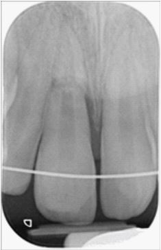
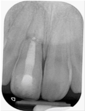
A 13-year-old male was referred to the Paediatric Dentistry Department, reporting a history of trauma on the maxillary right permanent central incisor (UR1). According to his clinical records, he was taken to a general dental practitioner, where the UR1 was splinted, and Amoxicillin was prescribed. When the patient came to our clinic, 20 days had passed, and the clinical examination showed soft tissue swelling related to the UR1 with mobility and a negative response to the pulp sensibility test. The radiographic evaluation confirmed a horizontal middle root fracture of the UR1 with the coronal fragment not approximated to the apical fragment and widening periodontal ligament space mesially (Figure 1).
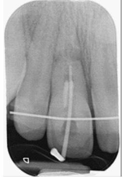
After obtaining informed consent from the patient and his parent, treatment was initiated under local anaesthesia. Following the isolation of the UR1 with a rubber dam, the access cavity was prepared. The contaminated canal remnants were removed, and the initial working length of 17 mm was obtained to the fracture line with size 30 K-file (Figure 2). The canal was irrigated with 2% sodium hypochlorite and dried with sterile paper points and non-sitting calcium hydroxide was selected for intracanal dressing and covered with sterile cotton pellets before sealing the coronal access with a Resin-Modified Glass ionomer Cement (RMGIC) filling. At this appointment, the splint and bulky composite were removed and replaced with a passive and flexible splint for improving oral hygiene.
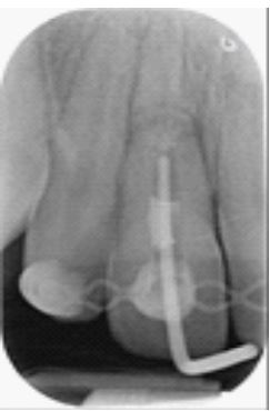
One month later, the patient returned. The UR1 was re-accessed, the cotton pellet was removed, and the canal was irrigated with 2% sodium hypochlorite. The canal was then instrumented to the fracture line with a size 80 K-file using a step-back technique and irrigated with 2% sodium hypochlorite. After drying the canal with sterile paper points, non-sitting calcium hydroxide was placed for intracanal dressing and covered with sterile cotton pellets before sealing the coronal access with a RMGIC filling. Two months later, the patient returned. The UR1 was re-accessed, the cotton pellet was removed, and the canal was irrigated with 2% sodium hypochlorite. The canal was then dried with sterile paper points, and a master apical cone size 80 was placed to the fracture line at the working length of 17 mm. A radiograph was taken to confirm the working length (Figure 3).
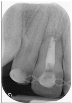
After that, the canal was obturated to the fracture line with TotalFill bioceramic putty and assessed by taking a radiograph (Figure 4). The cavity was cleaned and sealed with a RMGIC and a composite resin. The splint was removed at this visit.
A six-month review radiograph showed complete healing between the fragments, and the tooth was asymptomatic with no mobility (Figure 5).
Discussion
The management of horizontal root fractures is influenced by the stage of root formation, the degree of coronary fragment displacement, the time between trauma and treatment and the location of the fracture [8]. The incidence of pulp vitality is 96% when fractures are located in the apical third, 86% in the middle third and 20% in the cervical third [10]. An endodontic treatment is recommended when the pulp becomes necrotic [11].
The traumatized tooth must be followed up clinically and radiographically as the false negative response to pulp sensibility testing may be temporary for several months with fragment revascularization [5,11]. Because possible collateral circulation might occur in the fractured area, which helps maintain pulp vitality for a certain period [12]. If the pulp sensibility tests are still negative after three months and/or a radiographically radiolucent area appears between the fracture areas, an endodontic treatment is only required for the coronary fragment [8,13]. In the present case, the endodontic treatment for the coronary fragment was performed as the traumatized tooth was necrotic with the presence of swelling and a negative response to the pulp sensibility test 20 days after the trauma, leaving the apical fragment with no endodontic treatment due to its revascularization ability [8,13].
Due to the lack of apical constriction, sealing the coronary fragment might be more difficult. Calcium hydroxide has been used for stimulating the formation of apical plugs [8]. However, it requires many sessions to be changed; the root canal is at risk of reinfection and susceptibility to fracture of the treated roots [8]. Materials such as MTA (Mineral Trioxide Aggregate) have been used as an apical barrier due to their biological and chemical properties. The use of MTA materials is reduced because of the risk of teeth discoloration, complicated manipulation, longer sitting time, and the potential release of heavy metals [14]. Bioceramic materials have recently been introduced in the market, showing the benefits of bioceramic formulation, such as inducing tissue regeneration and antimicrobial properties. They are also ready-to-use presentations facilitating product removal for on-site preparation and simplifying this procedure which save time [15]. So, the selected material for the root canal filling in the present case was TotalFill bioceramic putty material. After six months of follow-up, the patient did not present painful symptoms or tooth discolouration, and the radiograph showed no lesion, which characterizes a successful treatment.
Conclusion
Early diagnosis and prompt treatment in managing root fractures result in a very good long-term prognosis. Treating middle root fractures in paediatric patients demands a delicate balance between preserving pulp vitality and achieving aesthetic restoration. This case report demonstrates that TotalFill Bioceramic putty can effectively achieve both objectives, leading to favourable clinical and radiographic outcomes in paediatric endodontics.
References
- Andreasen FM, Andreasen JO, Andersson MC. Textbook and Color Atlas of Traumatic Injuries to the Teeth. 4th ed. Oxford: Blackwell. 2007.
- Andreasen FM, Andreasen JO. Crown fractures. In Textbook and Color Atlas of Traumatic Injuries to the Teeth. 3rd ed. Copenhagen: Munksgaard. 1994.
- Birch R, Rock WP. The incidence of complications following root fracture in permanent anterior teeth. Br Dent J. 1986; 160: 119-22.
- Caliskan MK, Pehlivan Y. Prognosis of root-fractured permanent incisors. Endod Dent Traumatol. 1996; 12: 129-36.
- Bourguignon C, Cohenca N, Lauridsen E, Flores MT, O’Connell AC, Day PF, et al. International Association of Dental Traumatology guidelines for the management of traumatic dental injuries: 1. Fractures and luxations. Dental Traumatology. 2020; 36: 314-330.
- Girelli CFM, Lima CO, Lacerda MFLS, Coellho RG, Silveira FF, Nunes E. The importance of bioceramics and computed tomography. in the late clinical management of a horizontal root fracture: A case report. J Clin Exp Dent. 2020; 12: e514-e518.
- Toscano MA, Zacharczuk GA, López GE. Root fracture in the medium third: Treatment and 5 years follow up. Rev Assoc Odontol Argent. 2019; 107: 103-109.
- Roig M, Espona J, Mercadé M, Duran Sindreu F.Horizontal root fracture treated with MTA, a case report with a 10-year followup. Dental Traumatology. 2011; 27: 460-463.
- Kim D, Yue W, Yoon TC, Park SH, Kim E. Healing of Horizontal Intra-alveolar Root Fractures after Endodontic Treatment with Mineral Trioxide Aggregate. Journal of Endodontics. 2016; 42: 230-235.
- Cvek M, Tsilingaridis G, Andreasen JO. Survival of 534 incisors after intra-alveolar root fracture in patients aged 7-17 years. Dent Traumatol. 2008; 24: 379-87.
- Cvek M, Andreasen JO, Borum MK.Healing of 208 intra-alveolar root fractures in patients aged 7-17 years. Dent Traumatol. 2001; 17: 53-62.
- Soares AJ, Nagata JY, Lima TFR, Zaia AA. Management of horizontal root fracture report of two cases. Int J of Dent Clin. 2013; 5: 25-8.
- Flores MT, Andersson L, Andreasen JO, Bakland LK, Malmgren B, Barnett F, et al. Guidelines for the management of traumatic dental injuries. I. Fractures and luxations of permanent teeth. Dent Traumatol. 2007; 23: 66-71.
- López-García S, Lozano A, García-Bernal D, et. al. Biological effects of new hydraulic materials on human periodontal ligament stem cells. J. Clin. Med. 2019; 8: 12-16.
- Oliveira JCM, Silva FSB, Pinto SSL. Horizontal root fracture: case report. Revista Brasileira de Odontologia. 2008; 65: 76-79.